2YQC

Crystal Structure of uridine-diphospho-N-acetylglucosamine pyrophosphorylase from Candida albicans, in the apo-like form


Experimental Data Snapshot

  • Method: X-RAY DIFFRACTION
  • Resolution: 1.90 Å
  • R-Value Free: 0.201 
  • R-Value Work: 0.174 
  • R-Value Observed: 0.174 

wwPDB Validation   3D Report Full Report


This is version 1.4 of the entry. See complete history


Literature

Crystal Structure of Uridine-diphospho-N-acetylglucosamine Pyrophosphorylase from Candida albicans and Catalytic Reaction Mechanism

Maruyama, D.Nishitani, Y.Nonaka, T.Kita, A.Fukami, T.A.Mio, T.Yamada-Okabe, H.Yamada-Okabe, T.Miki, K.

(2007) J Biol Chem 282: 17221-17230

  • DOI: https://doi.org/10.1074/jbc.M611873200
  • Primary Citation of Related Structures:  
    2YQC, 2YQH, 2YQJ, 2YQS

  • PubMed Abstract: 

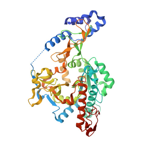
    Uridine-diphospho-N-acetylglucosamine (UDP-GlcNAc) is a precursor of the bacterial and fungal cell wall. It is also used in a component of N-linked glycosylation and the glycosylphosphoinositol anchor of eukaryotic proteins. It is synthesized from N-acetylglucosamine-1-phosphate (GlcNAc-1-P) and uridine-5'-triphosphate (UTP) by UDP-GlcNAc pyrophosphorylase (UAP). This is an S(N)2 reaction; the non-esterified oxygen atom of the GlcNAc-1-P phosphate group attacks the alpha-phosphate group of UTP. We determined crystal structures of UAP from Candida albicans (CaUAP1) without any ligands and also complexed with its substrate or with its product. The series of structures in different forms shows the induced fit movements of CaUAP1. Three loops approaching the ligand molecule close the active site when ligand is bound. In addition, Lys-421, instead of the metal ion in prokaryotic UAPs, is coordinated by both phosphate groups of UDP-Glc-NAc and acts as a cofactor. However, a magnesium ion enhances the enzymatic activity of CaUAP1, and thus we propose that the magnesium ion increases the affinity between UTP and the enzyme by coordinating to the alpha- and gamma-phosphate group of UTP.


  • Organizational Affiliation

    Department of Chemistry, Graduate School of Science, Kyoto University, Sakyo-ku, Kyoto 606-8502, Japan.


Macromolecules
Find similar proteins by:  (by identity cutoff)  |  3D Structure
Entity ID: 1
MoleculeChains Sequence LengthOrganismDetailsImage
UDP-N-acetylglucosamine pyrophosphorylase486Candida albicansMutation(s): 1 
EC: 2.7.7.23
UniProt
Find proteins for O74933 (Candida albicans)
Explore O74933 
Go to UniProtKB:  O74933
Entity Groups  
Sequence Clusters30% Identity50% Identity70% Identity90% Identity95% Identity100% Identity
UniProt GroupO74933
Sequence Annotations
Expand
  • Reference Sequence
Experimental Data & Validation

Experimental Data

  • Method: X-RAY DIFFRACTION
  • Resolution: 1.90 Å
  • R-Value Free: 0.201 
  • R-Value Work: 0.174 
  • R-Value Observed: 0.174 
  • Space Group: P 1 21 1
Unit Cell:
Length ( Å )Angle ( ˚ )
a = 47.774α = 90
b = 62.891β = 97.82
c = 90.602γ = 90
Software Package:
Software NamePurpose
CNSrefinement
HKL-2000data collection
HKL-2000data reduction
SCALEPACKdata scaling
AMoREphasing

Structure Validation

View Full Validation Report


Entry History 

Deposition Data

Revision History  (Full details and data files)

  • Version 1.0: 2007-05-22
    Type: Initial release
  • Version 1.1: 2008-04-30
    Changes: Version format compliance
  • Version 1.2: 2011-07-13
    Changes: Non-polymer description, Version format compliance
  • Version 1.3: 2021-11-10
    Changes: Database references, Derived calculations
  • Version 1.4: 2023-10-25
    Changes: Data collection, Refinement description