2Y9M

Pex4p-Pex22p structure


Experimental Data Snapshot

  • Method: X-RAY DIFFRACTION
  • Resolution: 2.60 Å
  • R-Value Free: 0.259 
  • R-Value Work: 0.205 
  • R-Value Observed: 0.208 

wwPDB Validation   3D Report Full Report


This is version 1.5 of the entry. See complete history


Literature

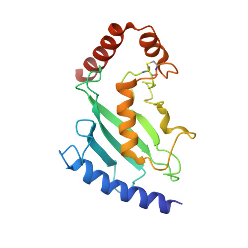
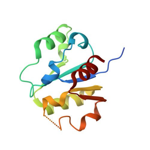
Insights Into Ubiquitin-Conjugating Enzyme/ Co-Activator Interactions from the Structure of the Pex4P:Pex22P Complex.

Williams, C.van den Berg, M.Panjikar, S.Stanley, W.A.Distel, B.Wilmanns, M.

(2011) EMBO J 31: 391

  • DOI: https://doi.org/10.1038/emboj.2011.411
  • Primary Citation of Related Structures:  
    2Y9M

  • PubMed Abstract: 

    Ubiquitin-conjugating enzymes (E2s) coordinate distinct types of ubiquitination via specific E3 ligases, to a large number of protein substrates. While many E2 enzymes need only the presence of an E3 ligase for substrate ubiquitination, a number of E2s require additional, non-canonical binding partners to specify their function. Here, we have determined the crystal structure and function of an E2/co-activator assembly, the Pex4p:Pex22p complex. The peroxisome-associated E2 enzyme Pex4p binds the peroxisomal membrane protein Pex22p through a binding site that does not overlap with any other known interaction interface in E2 enzymes. Pex22p association enhances Pex4p's ability to transfer ubiquitin to a substrate in vitro, and Pex22p binding-deficient forms of Pex4p are unable to ubiquitinate the peroxisomal import receptor Pex5p in vivo. Our data demonstrate that the Pex4p:Pex22p assembly, and not Pex4p alone, functions as the E2 enzyme required for Pex5p ubiquitination, establishing a novel mechanism of E2 enzyme regulation.


  • Organizational Affiliation

    Structural Biology Unit, European Molecular Biology Laboratory, Hamburg, Germany. c.williams@embl-hamburg.de


Macromolecules
Find similar proteins by:  (by identity cutoff)  |  3D Structure
Entity ID: 1
MoleculeChains Sequence LengthOrganismDetailsImage
UBIQUITIN-CONJUGATING ENZYME E2-21 KDA172Saccharomyces cerevisiaeMutation(s): 1 
EC: 6.3.2.19
UniProt
Find proteins for P29340 (Saccharomyces cerevisiae (strain ATCC 204508 / S288c))
Explore P29340 
Go to UniProtKB:  P29340
Entity Groups  
Sequence Clusters30% Identity50% Identity70% Identity90% Identity95% Identity100% Identity
UniProt GroupP29340
Sequence Annotations
Expand
  • Reference Sequence
Find similar proteins by:  (by identity cutoff)  |  3D Structure
Entity ID: 2
MoleculeChains Sequence LengthOrganismDetailsImage
PEROXISOME ASSEMBLY PROTEIN 22130Saccharomyces cerevisiaeMutation(s): 0 
UniProt
Find proteins for P39718 (Saccharomyces cerevisiae (strain ATCC 204508 / S288c))
Explore P39718 
Go to UniProtKB:  P39718
Entity Groups  
Sequence Clusters30% Identity50% Identity70% Identity90% Identity95% Identity100% Identity
UniProt GroupP39718
Sequence Annotations
Expand
  • Reference Sequence
Experimental Data & Validation

Experimental Data

  • Method: X-RAY DIFFRACTION
  • Resolution: 2.60 Å
  • R-Value Free: 0.259 
  • R-Value Work: 0.205 
  • R-Value Observed: 0.208 
  • Space Group: C 1 2 1
Unit Cell:
Length ( Å )Angle ( ˚ )
a = 139.328α = 90
b = 43.194β = 100.62
c = 60.474γ = 90
Software Package:
Software NamePurpose
XDSdata reduction
SCALAdata scaling
Auto-Rickshawphasing
autoSHARPphasing
REFMACrefinement

Structure Validation

View Full Validation Report


Entry History 

Deposition Data

Revision History  (Full details and data files)

  • Version 1.0: 2011-10-26
    Type: Initial release
  • Version 1.1: 2011-11-23
    Changes: Database references
  • Version 1.2: 2011-12-07
    Changes: Database references
  • Version 1.3: 2012-01-25
    Changes: Other
  • Version 1.4: 2012-04-18
    Changes: Other
  • Version 1.5: 2017-12-13
    Changes: Database references