2XNS

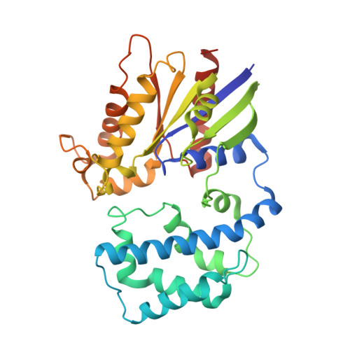
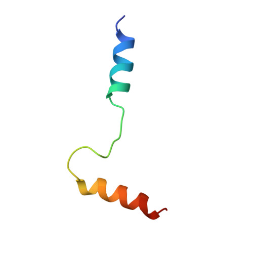
Crystal Structure Of Human G alpha i1 Bound To A Designed Helical Peptide Derived From The Goloco Motif Of RGS14


Experimental Data Snapshot

  • Method: X-RAY DIFFRACTION
  • Resolution: 3.41 Å
  • R-Value Free: 0.244 
  • R-Value Work: 0.223 
  • R-Value Observed: 0.223 

wwPDB Validation   3D Report Full Report


Ligand Structure Quality Assessment 


This is version 1.3 of the entry. See complete history


Literature

Computational Design of the Sequence and Structure of a Protein-Binding Peptide.

Sammond, D.W.Bosch, D.E.Butterfoss, G.L.Purbeck, C.Machius, M.Siderovski, D.P.Kuhlman, B.

(2011) J Am Chem Soc 133: 4190

  • DOI: https://doi.org/10.1021/ja110296z
  • Primary Citation of Related Structures:  
    2XNS

  • PubMed Abstract: 

    The de novo design of protein-binding peptides is challenging because it requires the identification of both a sequence and a backbone conformation favorable for binding. We used a computational strategy that iterates between structure and sequence optimization to redesign the C-terminal portion of the RGS14 GoLoco motif peptide so that it adopts a new conformation when bound to Gα(i1). An X-ray crystal structure of the redesigned complex closely matches the computational model, with a backbone root-mean-square deviation of 1.1 Å.


  • Organizational Affiliation

    Department of Biochemistry and Biophysics, University of North Carolina, Chapel Hill, North Carolina 27599-7260, USA.


Macromolecules
Find similar proteins by:  (by identity cutoff)  |  3D Structure
Entity ID: 1
MoleculeChains Sequence LengthOrganismDetailsImage
GUANINE NUCLEOTIDE-BINDING PROTEIN G(I) SUBUNIT ALPHA-1
A, B
327Homo sapiensMutation(s): 0 
EC: 3.6.5.1
UniProt & NIH Common Fund Data Resources
Find proteins for P63096 (Homo sapiens)
Explore P63096 
Go to UniProtKB:  P63096
PHAROS:  P63096
GTEx:  ENSG00000127955 
Entity Groups  
Sequence Clusters30% Identity50% Identity70% Identity90% Identity95% Identity100% Identity
UniProt GroupP63096
Sequence Annotations
Expand
  • Reference Sequence
Find similar proteins by:  (by identity cutoff)  |  3D Structure
Entity ID: 2
MoleculeChains Sequence LengthOrganismDetailsImage
REGULATOR OF G-PROTEIN SIGNALING 14
C, D
40Homo sapiensMutation(s): 0 
UniProt & NIH Common Fund Data Resources
Find proteins for O43566 (Homo sapiens)
Explore O43566 
Go to UniProtKB:  O43566
PHAROS:  O43566
GTEx:  ENSG00000169220 
Entity Groups  
Sequence Clusters30% Identity50% Identity70% Identity90% Identity95% Identity100% Identity
UniProt GroupO43566
Sequence Annotations
Expand
  • Reference Sequence
Experimental Data & Validation

Experimental Data

  • Method: X-RAY DIFFRACTION
  • Resolution: 3.41 Å
  • R-Value Free: 0.244 
  • R-Value Work: 0.223 
  • R-Value Observed: 0.223 
  • Space Group: I 2 3
Unit Cell:
Length ( Å )Angle ( ˚ )
a = 265.261α = 90
b = 265.261β = 90
c = 265.261γ = 90
Software Package:
Software NamePurpose
REFMACrefinement
HKL-2000data reduction
HKL-2000data scaling
PHASERphasing

Structure Validation

View Full Validation Report


Ligand Structure Quality Assessment 


Entry History 

Deposition Data

Revision History  (Full details and data files)

  • Version 1.0: 2011-06-08
    Type: Initial release
  • Version 1.1: 2011-07-13
    Changes: Version format compliance
  • Version 1.2: 2019-05-08
    Changes: Data collection, Experimental preparation, Other
  • Version 1.3: 2023-12-20
    Changes: Data collection, Database references, Derived calculations, Other, Refinement description