2WN7

Structural Basis for Substrate Recognition in the Enzymatic Component of ADP-ribosyltransferase Toxin CDTa from Clostridium difficile


Experimental Data Snapshot

  • Method: X-RAY DIFFRACTION
  • Resolution: 2.25 Å
  • R-Value Free: 0.265 
  • R-Value Work: 0.205 
  • R-Value Observed: 0.208 

wwPDB Validation   3D Report Full Report


Ligand Structure Quality Assessment 


This is version 1.3 of the entry. See complete history


Literature

Structural Basis for Substrate Recognition in the Enzymatic Component of Adp-Ribosyltransferase Toxin Cdta from Clostridium Difficile.

Sundriyal, A.Roberts, A.K.Shone, C.C.Acharya, K.R.

(2009) J Biol Chem 284: 28713

  • DOI: https://doi.org/10.1074/jbc.M109.043018
  • Primary Citation of Related Structures:  
    2WN4, 2WN5, 2WN6, 2WN7, 2WN8

  • PubMed Abstract: 

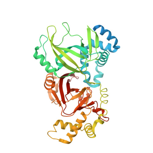
    ADP-ribosylation is one of the favored modes of cell intoxication employed by several bacteria. Clostridium difficile is recognized to be an important nosocomial pathogen associated with considerable morbidity and attributable mortality. Along with its two well known toxins, Toxin A and Toxin B, it produces an ADP-ribosylating toxin that targets monomeric actin of the target cell. Like other Clostridial actin ADP-ribosylating toxins, this binary toxin, known as C. difficile toxin (CDT), is composed of two subunits, CDTa and CDTb. In this study, we present high resolution crystal structures of CDTa in its native form (at pH 4.0, 8.5, and 9.0) and in complex with ADP-ribose donors, NAD and NADPH (at pH 9.0). The crystal structures of the native protein show "pronounced conformational flexibility" confined to the active site region of the protein and "enhanced" disorder at low pH, whereas the complex structures highlight significant differences in "ligand specificity" compared with the enzymatic subunit of a close homologue, Clostridium perfringens iota toxin. Specifically in CDTa, two of the suggested catalytically important residues (Glu-385 and Glu-387) seem to play no role or a less important role in ligand binding. These structural data provide the first detailed information on protein-donor substrate complex stabilization in CDTa, which may have implications in understanding CDT recognition.


  • Organizational Affiliation

    Department of Biology and Biochemistry, University of Bath, Claverton Down, Bath BA2 7AY, United Kingdom.


Macromolecules
Find similar proteins by:  (by identity cutoff)  |  3D Structure
Entity ID: 1
MoleculeChains Sequence LengthOrganismDetailsImage
ADP-RIBOSYLTRANSFERASE ENZYMATIC COMPONENT463Clostridioides difficileMutation(s): 0 
UniProt
Find proteins for Q9KH42 (Clostridioides difficile)
Explore Q9KH42 
Go to UniProtKB:  Q9KH42
Entity Groups  
Sequence Clusters30% Identity50% Identity70% Identity90% Identity95% Identity100% Identity
UniProt GroupQ9KH42
Sequence Annotations
Expand
  • Reference Sequence
Small Molecules
Ligands 2 Unique
IDChains Name / Formula / InChI Key2D Diagram3D Interactions
NAD
Query on NAD

Download Ideal Coordinates CCD File 
B [auth A]NICOTINAMIDE-ADENINE-DINUCLEOTIDE
C21 H27 N7 O14 P2
BAWFJGJZGIEFAR-NNYOXOHSSA-N
GOL
Query on GOL

Download Ideal Coordinates CCD File 
C [auth A]GLYCEROL
C3 H8 O3
PEDCQBHIVMGVHV-UHFFFAOYSA-N
Experimental Data & Validation

Experimental Data

  • Method: X-RAY DIFFRACTION
  • Resolution: 2.25 Å
  • R-Value Free: 0.265 
  • R-Value Work: 0.205 
  • R-Value Observed: 0.208 
  • Space Group: P 1 21 1
Unit Cell:
Length ( Å )Angle ( ˚ )
a = 62.057α = 90
b = 46.772β = 97.73
c = 77.722γ = 90
Software Package:
Software NamePurpose
REFMACrefinement
HKL-2000data reduction
HKL-2000data scaling
MOLREPphasing

Structure Validation

View Full Validation Report


Ligand Structure Quality Assessment 


Entry History 

Deposition Data

Revision History  (Full details and data files)

  • Version 1.0: 2009-08-18
    Type: Initial release
  • Version 1.1: 2011-05-08
    Changes: Version format compliance
  • Version 1.2: 2011-07-13
    Changes: Version format compliance
  • Version 1.3: 2023-12-13
    Changes: Data collection, Database references, Derived calculations, Other, Refinement description