2VOD

Crystal structure of N-terminal domains of Human La protein complexed with RNA oligomer AUAUUUU


Experimental Data Snapshot

  • Method: X-RAY DIFFRACTION
  • Resolution: 2.10 Å
  • R-Value Free: 0.272 
  • R-Value Work: 0.232 
  • R-Value Observed: 0.232 

wwPDB Validation   3D Report Full Report


This is version 1.2 of the entry. See complete history


Literature

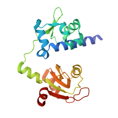
Structural Analysis Reveals Conformational Plasticity in the Recognition of RNA 3' Ends by the Human La Protein.

Kotik-Kogan, O.Valentine, E.R.Sanfelice, D.Conte, M.R.Curry, S.

(2008) Structure 16: 852

  • DOI: https://doi.org/10.1016/j.str.2008.02.021
  • Primary Citation of Related Structures:  
    2VOD, 2VON, 2VOO, 2VOP

  • PubMed Abstract: 

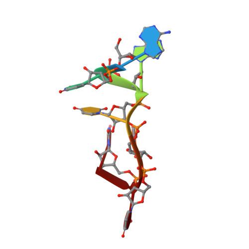
    The eukaryotic La protein recognizes the 3' poly(U) sequences of nascent RNA polymerase III transcripts to assist folding and maturation. The 3' ends of such RNAs are bound by the N-terminal domain of La (LaNTD). We have solved the crystal structures of four LaNTD:RNA complexes, each containing a different single-stranded RNA oligomer, and compared them to the structure of a previously published LaNTD:RNA complex containing partially duplex RNA. The presence of purely single-stranded RNA in the binding pocket at the interface between the La motif and RRM domains allows significantly closer contact with the 3' end of the RNA. Comparison of the different LaNTD:RNA complexes identifies a conserved set of interactions with the last two nucleotides at the 3' end of the RNA ligand that are key to binding. Strikingly, we also observe two alternative conformations of bound ssRNA, indicative of an unexpected degree of plasticity in the modes of RNA binding.


  • Organizational Affiliation

    Biophysics Section, Blackett Laboratory, Imperial College, Exhibition Road, London, United Kingdom.


Macromolecules

Find similar proteins by:  (by identity cutoff)  |  3D Structure
Entity ID: 1
MoleculeChains Sequence LengthOrganismDetailsImage
LUPUS LA PROTEIN
A, B
193Homo sapiensMutation(s): 0 
UniProt & NIH Common Fund Data Resources
Find proteins for P05455 (Homo sapiens)
Explore P05455 
Go to UniProtKB:  P05455
PHAROS:  P05455
GTEx:  ENSG00000138385 
Entity Groups  
Sequence Clusters30% Identity50% Identity70% Identity90% Identity95% Identity100% Identity
UniProt GroupP05455
Sequence Annotations
Expand
  • Reference Sequence

Find similar nucleic acids by:  Sequence   |   3D Structure  

Entity ID: 2
MoleculeChains LengthOrganismImage
5'-R(*AP*UP*AP*UP*UP*UP*UP)-3'
C, D
7Homo sapiens
Sequence Annotations
Expand
  • Reference Sequence
Experimental Data & Validation

Experimental Data

  • Method: X-RAY DIFFRACTION
  • Resolution: 2.10 Å
  • R-Value Free: 0.272 
  • R-Value Work: 0.232 
  • R-Value Observed: 0.232 
  • Space Group: C 1 2 1
Unit Cell:
Length ( Å )Angle ( ˚ )
a = 140.029α = 90
b = 44.471β = 114.35
c = 91.285γ = 90
Software Package:
Software NamePurpose
CNSrefinement
MOSFLMdata reduction
SCALAdata scaling
PHASERphasing

Structure Validation

View Full Validation Report


Entry History 

Deposition Data

Revision History  (Full details and data files)

  • Version 1.0: 2008-05-06
    Type: Initial release
  • Version 1.1: 2011-05-08
    Changes: Version format compliance
  • Version 1.2: 2011-07-13
    Changes: Version format compliance