2QVB

Crystal Structure of Haloalkane Dehalogenase Rv2579 from Mycobacterium tuberculosis


Experimental Data Snapshot

  • Method: X-RAY DIFFRACTION
  • Resolution: 1.19 Å
  • R-Value Free: 0.160 
  • R-Value Work: 0.120 

wwPDB Validation   3D Report Full Report


This is version 1.3 of the entry. See complete history


Literature

X-ray crystal structure of Mycobacterium tuberculosis haloalkane dehalogenase Rv2579.

Mazumdar, P.A.Hulecki, J.C.Cherney, M.M.Garen, C.R.James, M.N.

(2008) Biochim Biophys Acta 1784: 351-362

  • DOI: https://doi.org/10.1016/j.bbapap.2007.10.014
  • Primary Citation of Related Structures:  
    2QVB

  • PubMed Abstract: 

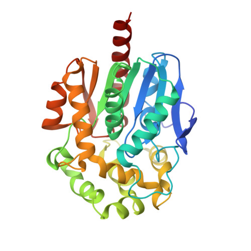
    Haloalkane dehalogenases are enzymes well known to be important in bioremediation; the organisms from which they are produced are able to clean up toxic organohalides from polluted environments. However, besides being found in such contaminated environments, these enzymes have also been found in root or tissue-colonizing bacterial species. The haloalkane dehalogenase Rv2579 from Mycobacterium tuberculosis H37Rv has been cloned, expressed, purified and its crystal structure determined at high resolution (1.2A). In addition, the crystal structure of the enzyme has been determined in complex with the product from the reaction with 1,3-dibromopropane, i.e. 1,3-propanediol and in complex with the classical substrate of haloalkane dehalogenases, 1,2-dichloroethane. The enzyme is a two-domain protein having a catalytic domain of an alpha/beta hydrolase fold and a cap domain. The active site residues and the halide-stabilizing residues have been identified as Asp109, Glu133, His273, Asn39 and Trp110. Its overall structure is similar to those of other known haloalkane dehalogenases. Its mechanism of action involves an SN2 nucleophilic displacement.


  • Organizational Affiliation

    Group in Protein Structure and Function, Department of Biochemistry, University of Alberta, Edmonton, Alberta, Canada T6G 2H7.


Macromolecules
Find similar proteins by:  (by identity cutoff)  |  3D Structure
Entity ID: 1
MoleculeChains Sequence LengthOrganismDetailsImage
Haloalkane dehalogenase 3
A, B
297Mycobacterium tuberculosis H37RvMutation(s): 0 
Gene Names: dhaA
EC: 3.8.1.5
UniProt
Find proteins for P9WMR9 (Mycobacterium tuberculosis (strain ATCC 25618 / H37Rv))
Explore P9WMR9 
Go to UniProtKB:  P9WMR9
Entity Groups  
Sequence Clusters30% Identity50% Identity70% Identity90% Identity95% Identity100% Identity
UniProt GroupP9WMR9
Sequence Annotations
Expand
  • Reference Sequence
Experimental Data & Validation

Experimental Data

  • Method: X-RAY DIFFRACTION
  • Resolution: 1.19 Å
  • R-Value Free: 0.160 
  • R-Value Work: 0.120 
  • Space Group: P 21 21 21
Unit Cell:
Length ( Å )Angle ( ˚ )
a = 60.7α = 90
b = 65.7β = 90
c = 129.7γ = 90
Software Package:
Software NamePurpose
DENZOdata reduction
SCALEPACKdata scaling
REFMACrefinement
PDB_EXTRACTdata extraction
ADSCdata collection
HKL-2000data reduction
PHASERphasing

Structure Validation

View Full Validation Report


Entry History 

Deposition Data

Revision History  (Full details and data files)

  • Version 1.0: 2008-02-12
    Type: Initial release
  • Version 1.1: 2011-07-13
    Changes: Source and taxonomy, Version format compliance
  • Version 1.2: 2017-10-25
    Changes: Advisory, Refinement description
  • Version 1.3: 2023-08-30
    Changes: Advisory, Data collection, Database references, Derived calculations, Refinement description