


2QT9
Human dipeptidyl peptidase iv/cd26 in complex with a 4-aryl cyclohexylalanine inhibitor
- PDB DOI: https://doi.org/10.2210/pdb2QT9/pdb
- Classification: HYDROLASE
- Organism(s): Homo sapiens
- Expression System: Spodoptera frugiperda
- Mutation(s): Yes
- Deposited: 2007-08-01 Released: 2007-11-06
Experimental Data Snapshot
- Method: X-RAY DIFFRACTION
- Resolution: 2.10 Å
- R-Value Free: 0.229
- R-Value Work: 0.190

This is version 2.2 of the entry. See complete history.

Macromolecules
Find similar proteins by:
(by identity cutoff) | 3D Structure
Entity ID: 1 | |||||
|---|---|---|---|---|---|
| Molecule | Chains | Sequence Length | Organism | Details | Image |
| Dipeptidyl peptidase 4 | 766 | Homo sapiens | Mutation(s): 1 Gene Names: DPP4, ADCP2, CD26 EC: 3.4.14.5 | 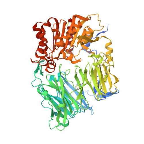 | |
UniProt & NIH Common Fund Data Resources | |||||
Find proteins for P27487 (Homo sapiens) Explore P27487 Go to UniProtKB: P27487 | |||||
PHAROS: P27487 GTEx: ENSG00000197635 | |||||
Entity Groups | |||||
| Sequence Clusters | 30% Identity50% Identity70% Identity90% Identity95% Identity100% Identity | ||||
| UniProt Group | P27487 | ||||
Sequence AnnotationsExpand | |||||
| |||||
Oligosaccharides
Entity ID: 2 | |||||
|---|---|---|---|---|---|
| Molecule | Chains | Length | 2D Diagram | Glycosylation | 3D Interactions |
| 2-acetamido-2-deoxy-alpha-D-glucopyranose-(1-4)-2-acetamido-2-deoxy-beta-D-glucopyranose | C [auth L], F [auth O] | 2 |  | N-Glycosylation | |
Glycosylation Resources | |||||
GlyTouCan: G07375KG GlyCosmos: G07375KG GlyGen: G07375KG | |||||
Entity ID: 3 | |||||
|---|---|---|---|---|---|
| Molecule | Chains | Length | 2D Diagram | Glycosylation | 3D Interactions |
| 2-acetamido-2-deoxy-beta-D-glucopyranose-(1-4)-2-acetamido-2-deoxy-beta-D-glucopyranose | D [auth M], E [auth N], G [auth P], H [auth Q], I [auth R], D [auth M], E [auth N], G [auth P], H [auth Q], I [auth R], J [auth S], K [auth T] | 2 |  | N-Glycosylation | |
Glycosylation Resources | |||||
GlyTouCan: G42666HT GlyCosmos: G42666HT GlyGen: G42666HT | |||||
Small Molecules
| Ligands 3 Unique | |||||
|---|---|---|---|---|---|
| ID | Chains | Name / Formula / InChI Key | 2D Diagram | 3D Interactions | |
| 524 Query on 524 | P [auth A], T [auth B] | (2S,3S)-3-AMINO-4-[(3S)-3-FLUOROPYRROLIDIN-1-YL]-N,N-DIMETHYL-4-OXO-2-(TRANS-4-[1,2,4]TRIAZOLO[1,5-A]PYRIDIN-5-YLCYCLOH
EXYL)BUTANAMIDE C22 H31 F N6 O2 ZPWDKZWKUOYOHA-UKSSEWCLSA-N |  | ||
| NAG Query on NAG | L [auth A] M [auth A] N [auth A] Q [auth B] R [auth B] | 2-acetamido-2-deoxy-beta-D-glucopyranose C8 H15 N O6 OVRNDRQMDRJTHS-FMDGEEDCSA-N |  | ||
| NA Query on NA | O [auth A] | SODIUM ION Na FKNQFGJONOIPTF-UHFFFAOYSA-N |  | ||
Experimental Data & Validation
Experimental Data
- Method: X-RAY DIFFRACTION
- Resolution: 2.10 Å
- R-Value Free: 0.229
- R-Value Work: 0.190
- Space Group: P 21 21 21
Unit Cell:
| Length ( Å ) | Angle ( ˚ ) |
|---|---|
| a = 117.697 | α = 90 |
| b = 126.072 | β = 90 |
| c = 136.97 | γ = 90 |
| Software Name | Purpose |
|---|---|
| CNS | refinement |
| PDB_EXTRACT | data extraction |
| ADSC | data collection |
| HKL-2000 | data reduction |
| HKL-2000 | data scaling |
| CNX | phasing |
| CNX | refinement |
Entry History
Deposition Data
- Released Date: 2007-11-06 Deposition Author(s): Scapin, G.
Revision History (Full details and data files)
- Version 1.0: 2007-11-06
Type: Initial release - Version 1.1: 2011-07-13
Changes: Non-polymer description, Version format compliance - Version 2.0: 2020-07-29
Type: Remediation
Reason: Carbohydrate remediation
Changes: Advisory, Atomic model, Data collection, Derived calculations, Structure summary - Version 2.1: 2021-10-20
Changes: Database references, Structure summary - Version 2.2: 2023-08-30
Changes: Data collection, Refinement description
