2PO0

Crystal structure of the P. abyssi exosome RNase PH ring complexed with ADP in double conformation


Experimental Data Snapshot

  • Method: X-RAY DIFFRACTION
  • Resolution: 2.30 Å
  • R-Value Free: 0.243 
  • R-Value Work: 0.190 
  • R-Value Observed: 0.193 

wwPDB Validation   3D Report Full Report


Ligand Structure Quality Assessment 


This is version 1.2 of the entry. See complete history


Literature

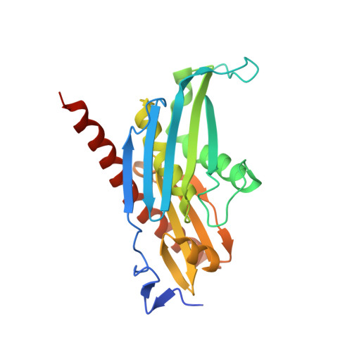
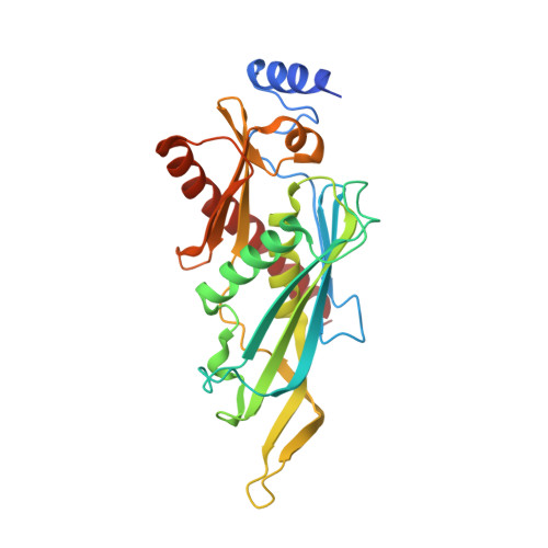
Insights into the mechanism of progressive RNA degradation by the archaeal exosome.

Navarro, M.V.A.S.Oliveira, C.C.Zanchin, N.I.Guimaraes, B.G.

(2008) J Biol Chem 283: 14120-14131

  • DOI: https://doi.org/10.1074/jbc.M801005200
  • Primary Citation of Related Structures:  
    2PNZ, 2PO0, 2PO1, 2PO2

  • PubMed Abstract: 

    Initially identified in yeast, the exosome has emerged as a central component of the RNA maturation and degradation machinery both in Archaea and eukaryotes. Here we describe a series of high-resolution structures of the RNase PH ring from the Pyrococcus abyssi exosome, one of them containing three 10-mer RNA strands within the exosome catalytic chamber, and report additional nucleotide interactions involving positions N5 and N7. Residues from all three Rrp41-Rrp42 heterodimers interact with a single RNA molecule, providing evidence for the functional relevance of exosome ring-like assembly in RNA processivity. Furthermore, an ADP-bound structure showed a rearrangement of nucleotide interactions at site N1, suggesting a rationale for the elimination of nucleoside diphosphate after catalysis. In combination with RNA degradation assays performed with mutants of key amino acid residues, the structural data presented here provide support for a model of exosome-mediated RNA degradation that integrates the events involving catalytic cleavage, product elimination, and RNA translocation. Finally, comparisons between the archaeal and human exosome structures provide a possible explanation for the eukaryotic exosome inability to catalyze phosphate-dependent RNA degradation.


  • Organizational Affiliation

    Brazilian Synchrotron Light Laboratory, 13083-970 Campinas, SP, Brazil. mn372@cornell.edu


Macromolecules
Find similar proteins by:  (by identity cutoff)  |  3D Structure
Entity ID: 1
MoleculeChains Sequence LengthOrganismDetailsImage
Probable exosome complex exonuclease 1249Pyrococcus abyssiMutation(s): 0 
Gene Names: Rrp41
EC: 3.1.13
UniProt
Find proteins for Q9V119 (Pyrococcus abyssi (strain GE5 / Orsay))
Explore Q9V119 
Go to UniProtKB:  Q9V119
Entity Groups  
Sequence Clusters30% Identity50% Identity70% Identity90% Identity95% Identity100% Identity
UniProt GroupQ9V119
Sequence Annotations
Expand
  • Reference Sequence
Find similar proteins by:  (by identity cutoff)  |  3D Structure
Entity ID: 2
MoleculeChains Sequence LengthOrganismDetailsImage
Probable exosome complex exonuclease 2277Pyrococcus abyssiMutation(s): 0 
Gene Names: Rrp42
EC: 3.1.13
UniProt
Find proteins for Q9V118 (Pyrococcus abyssi (strain GE5 / Orsay))
Explore Q9V118 
Go to UniProtKB:  Q9V118
Entity Groups  
Sequence Clusters30% Identity50% Identity70% Identity90% Identity95% Identity100% Identity
UniProt GroupQ9V118
Sequence Annotations
Expand
  • Reference Sequence
Experimental Data & Validation

Experimental Data

  • Method: X-RAY DIFFRACTION
  • Resolution: 2.30 Å
  • R-Value Free: 0.243 
  • R-Value Work: 0.190 
  • R-Value Observed: 0.193 
  • Space Group: P 3 2 1
Unit Cell:
Length ( Å )Angle ( ˚ )
a = 94.2α = 90
b = 94.2β = 90
c = 127.7γ = 120
Software Package:
Software NamePurpose
REFMACrefinement
MAR345dtbdata collection
XDSdata reduction
XDSdata scaling
MOLREPphasing

Structure Validation

View Full Validation Report


Ligand Structure Quality Assessment 


Entry History 

Deposition Data

Revision History  (Full details and data files)

  • Version 1.0: 2008-03-18
    Type: Initial release
  • Version 1.1: 2011-07-13
    Changes: Version format compliance
  • Version 1.2: 2023-08-30
    Changes: Data collection, Database references, Derived calculations, Refinement description