Crystal structure of a novel myotoxic Arg49 phospholipase A(2) homolog (zhaoermiatoxin) from Zhaoermia mangshanensis snake venom: Insights into Arg49 coordination and the role of Lys122 in the polarization of the C-terminus.
Murakami, M.T., Kuch, U., Betzel, C., Mebs, D., Arni, R.K.(2008) Toxicon 51: 723-735
- PubMed: 18295812
- DOI: https://doi.org/10.1016/j.toxicon.2007.11.018
- Primary Citation of Related Structures:
2PH4 - PubMed Abstract:
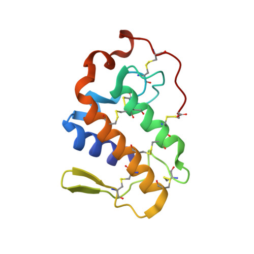
The venom of Zhaoermia mangshanensis, encountered solely in Mt Mang in China's Hunan Province, exhibits coagulant, phosphodiesterase, l-amino acid oxidase, kallikrein, phospholipase A2 and myotoxic activities. The catalytically inactive PLA2 homolog referred to as zhaoermiatoxin is highly myotoxic and displays high myonecrotic and edema activities. Zhaoermiatoxin possesses a molecular weight of 13,972Da, consists of 121 amino-acid residues cross-linked by seven disulfide bridges and shares high sequence homology with Lys49-PLA2s from the distantly related Asian pitvipers. However, zhaoermiatoxin possesses an arginine residue at position 49 instead of a lysine, thereby suggesting a secondary Lys49-->Arg substitution which results in a catalytically inactive protein. We have determined the first crystal structure of zhaoermiatoxin, an Arg49-PLA2, from Zhaoermia mangshanensis venom at 2.05 angstroms resolution, which represents a novel member of phospholipase A2 family. In this structure, unlike the Lys49 PLA2s, the C-terminus is well ordered and an unexpected non-polarized state of the putative calcium-binding loop due to the flip of Lys122 towards the bulk solvent is observed. The orientation of the Arg-49 side chain results in a similar binding mode to that observed in the Lys49 PLA2s; however, the guadinidium group is tri-coordinated by carbonyl oxygen atoms of the putative calcium-binding loop, whereas the Nzeta atom of lysine is tetra-coordinated as a result of the different conformation adopted by the putative calcium-binding loop.
Organizational Affiliation:
Center for Structural & Molecular Biology, Department of Physics, IBILCE/UNESP, R. Cristovao Colombo 2265, São José do Rio Preto, São Paulo CEP 15054-000, Brazil.