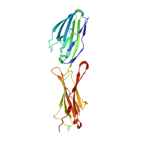
The Laminin 511/521-binding site on the Lutheran blood group glycoprotein is located at the flexible junction of Ig domains 2 and 3.
Mankelow, T.J., Burton, N., Stefansdottir, F.O., Spring, F.A., Parsons, S.F., Pedersen, J.S., Oliveira, C.L., Lammie, D., Wess, T., Mohandas, N., Chasis, J.A., Brady, R.L., Anstee, D.J.(2007) Blood 110: 3398-3406
- PubMed: 17638854
- DOI: https://doi.org/10.1182/blood-2007-06-094748
- Primary Citation of Related Structures:
2PET, 2PF6 - PubMed Abstract:
The Lutheran blood group glycoprotein, first discovered on erythrocytes, is widely expressed in human tissues. It is a ligand for the alpha5 subunit of Laminin 511/521, an extracellular matrix protein. This interaction may contribute to vaso-occlusive events that are an important cause of morbidity in sickle cell disease. Using x-ray crystallography, small-angle x-ray scattering, and site-directed mutagenesis, we show that the extracellular region of Lutheran forms an extended structure with a distinctive bend between the second and third immunoglobulin-like domains. The linker between domains 2 and 3 appears to be flexible and is a critical determinant in maintaining an overall conformation for Lutheran that is capable of binding to Laminin. Mutagenesis studies indicate that Asp312 of Lutheran and the surrounding cluster of negatively charged residues in this linker region form the Laminin-binding site. Unusually, receptor binding is therefore not a function of the domains expected to be furthermost from the plasma membrane. These studies imply that structural flexibility of Lutheran may be essential for its interaction with Laminin and present a novel opportunity for the development of therapeutics for sickle cell disease.
Organizational Affiliation:
Bristol Institute for Transfusion Sciences, National Blood Service, Southmead Road, Bristol, United Kingdom. tosti.mankelow@nbs.nhs.uk