2OYL

Endo-glycoceramidase II from Rhodococcus sp.: cellobiose-like imidazole complex


Experimental Data Snapshot

  • Method: X-RAY DIFFRACTION
  • Resolution: 1.80 Å
  • R-Value Free: 0.205 
  • R-Value Work: 0.174 
  • R-Value Observed: 0.175 

wwPDB Validation   3D Report Full Report


Ligand Structure Quality Assessment 


This is version 2.1 of the entry. See complete history


Literature

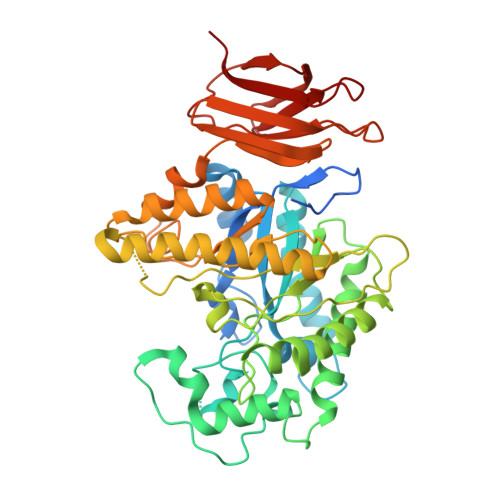
The structural basis of glycosidase inhibition by five-membered iminocyclitols: the clan a glycoside hydrolase endoglycoceramidase as a model system.

Caines, M.E.Hancock, S.M.Tarling, C.A.Wrodnigg, T.M.Stick, R.V.Stutz, A.E.Vasella, A.Withers, S.G.Strynadka, N.C.

(2007) Angew Chem Int Ed Engl 46: 4474-4476


Macromolecules
Find similar proteins by:  (by identity cutoff)  |  3D Structure
Entity ID: 1
MoleculeChains Sequence LengthOrganismDetailsImage
Endoglycoceramidase II
A, B
481Rhodococcus sp. (in: high G+C Gram-positive bacteria)Mutation(s): 0 
EC: 3.2.1.123
UniProt
Find proteins for O33853 (Rhodococcus sp)
Explore O33853 
Go to UniProtKB:  O33853
Entity Groups  
Sequence Clusters30% Identity50% Identity70% Identity90% Identity95% Identity100% Identity
UniProt GroupO33853
Sequence Annotations
Expand
  • Reference Sequence
Small Molecules
Ligands 3 Unique
IDChains Name / Formula / InChI Key2D Diagram3D Interactions
IDC
Query on IDC

Download Ideal Coordinates CCD File 
E [auth A],
J [auth B]
(5R,6R,7R,8S)-7,8-dihydroxy-5-(hydroxymethyl)-5,6,7,8-tetrahydroimidazo[1,2-a]pyridin-6-yl beta-D-glucopyranoside
C14 H22 N2 O9
CSXOUJBOYXGFCL-OFKZETBZSA-N
GOL
Query on GOL

Download Ideal Coordinates CCD File 
F [auth A],
G [auth A]
GLYCEROL
C3 H8 O3
PEDCQBHIVMGVHV-UHFFFAOYSA-N
NA
Query on NA

Download Ideal Coordinates CCD File 
C [auth A],
D [auth A],
H [auth B],
I [auth B]
SODIUM ION
Na
FKNQFGJONOIPTF-UHFFFAOYSA-N
Binding Affinity Annotations 
IDSourceBinding Affinity
IDC PDBBind:  2OYL Ki: 500 (nM) from 1 assay(s)
Binding MOAD:  2OYL Ki: 500 (nM) from 1 assay(s)
Experimental Data & Validation

Experimental Data

  • Method: X-RAY DIFFRACTION
  • Resolution: 1.80 Å
  • R-Value Free: 0.205 
  • R-Value Work: 0.174 
  • R-Value Observed: 0.175 
  • Space Group: P 1 21 1
Unit Cell:
Length ( Å )Angle ( ˚ )
a = 53.752α = 90
b = 93.246β = 98.35
c = 94.432γ = 90
Software Package:
Software NamePurpose
SCALAdata scaling
REFMACrefinement
PDB_EXTRACTdata extraction
MOSFLMdata reduction
CCP4data scaling

Structure Validation

View Full Validation Report


Ligand Structure Quality Assessment 


Entry History 

Deposition Data

Revision History  (Full details and data files)

  • Version 1.0: 2007-03-06
    Type: Initial release
  • Version 1.1: 2007-10-24
    Changes: Version format compliance
  • Version 1.2: 2011-07-13
    Changes: Non-polymer description, Version format compliance
  • Version 2.0: 2020-07-29
    Type: Remediation
    Reason: Carbohydrate remediation
    Changes: Advisory, Atomic model, Database references, Derived calculations, Structure summary
  • Version 2.1: 2023-08-30
    Changes: Advisory, Data collection, Database references, Refinement description, Structure summary