2OXN

Vibrio cholerae family 3 glycoside hydrolase (NagZ) in complex with PUGNAc


Experimental Data Snapshot

  • Method: X-RAY DIFFRACTION
  • Resolution: 1.70 Å
  • R-Value Free: 0.199 
  • R-Value Work: 0.168 
  • R-Value Observed: 0.170 

wwPDB Validation   3D Report Full Report


Ligand Structure Quality Assessment 


This is version 1.4 of the entry. See complete history


Literature

Small molecule inhibitors of a glycoside hydrolase attenuate inducible AmpC-mediated beta-lactam resistance.

Stubbs, K.A.Balcewich, M.Mark, B.L.Vocadlo, D.J.

(2007) J Biol Chem 282: 21382-21391

  • DOI: https://doi.org/10.1074/jbc.M700084200
  • Primary Citation of Related Structures:  
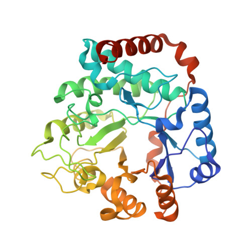
    2OXN

  • PubMed Abstract: 

    The increasing spread of plasmid-borne ampC-ampR operons is of considerable medical importance, since the AmpC beta-lactamases they encode confer high level resistance to many third generation cephalosporins. Induction of AmpC beta-lactamase from endogenous or plasmid-borne ampC-ampR operons is mediated by a catabolic inducer molecule, 1,6-anhydro-N-acetylmuramic acid (MurNAc) tripeptide, an intermediate of the cell wall recycling pathway derived from the peptidoglycan. Here we describe a strategy for attenuating the antibiotic resistance associated with the ampC-ampR operon by blocking the formation of the inducer molecule using small molecule inhibitors of NagZ, the glycoside hydrolase catalyzing the formation of this inducer molecule. The structure of the NagZ-inhibitor complex provides insight into the molecular basis for inhibition and enables the development of inhibitors with 100-fold selectivity for NagZ over functionally related human enzymes. These PUGNAc-derived inhibitors reduce the minimal inhibitory concentration (MIC) values for several clinically relevant cephalosporins in both wild-type and AmpC-hyperproducing strains lacking functional AmpD.


  • Organizational Affiliation

    Department of Chemistry, Simon Fraser University, Burnaby, British Columbia V5A 1S6.


Macromolecules
Find similar proteins by:  (by identity cutoff)  |  3D Structure
Entity ID: 1
MoleculeChains Sequence LengthOrganismDetailsImage
Beta-hexosaminidase340Vibrio choleraeMutation(s): 0 
Gene Names: nagZ
EC: 3.2.1.52
UniProt
Find proteins for Q9KU37 (Vibrio cholerae serotype O1 (strain ATCC 39315 / El Tor Inaba N16961))
Explore Q9KU37 
Go to UniProtKB:  Q9KU37
Entity Groups  
Sequence Clusters30% Identity50% Identity70% Identity90% Identity95% Identity100% Identity
UniProt GroupQ9KU37
Sequence Annotations
Expand
  • Reference Sequence
Small Molecules
Ligands 1 Unique
IDChains Name / Formula / InChI Key2D Diagram3D Interactions
OAN
Query on OAN

Download Ideal Coordinates CCD File 
B [auth A]O-(2-ACETAMIDO-2-DEOXY D-GLUCOPYRANOSYLIDENE) AMINO-N-PHENYLCARBAMATE
C15 H19 N3 O7
PBLNJFVQMUMOJY-JXZOILRNSA-N
Binding Affinity Annotations 
IDSourceBinding Affinity
OAN Binding MOAD:  2OXN Ki: 36 (nM) from 1 assay(s)
PDBBind:  2OXN Ki: 36 (nM) from 1 assay(s)
Experimental Data & Validation

Experimental Data

  • Method: X-RAY DIFFRACTION
  • Resolution: 1.70 Å
  • R-Value Free: 0.199 
  • R-Value Work: 0.168 
  • R-Value Observed: 0.170 
  • Space Group: P 21 21 21
Unit Cell:
Length ( Å )Angle ( ˚ )
a = 48.734α = 90
b = 67.85β = 90
c = 96.894γ = 90
Software Package:
Software NamePurpose
REFMACrefinement
PDB_EXTRACTdata extraction
CrystalCleardata collection
MOSFLMdata reduction
SCALAdata scaling
PHASERphasing

Structure Validation

View Full Validation Report


Ligand Structure Quality Assessment 


Entry History 

Deposition Data

Revision History  (Full details and data files)

  • Version 1.0: 2007-06-12
    Type: Initial release
  • Version 1.1: 2008-05-01
    Changes: Version format compliance
  • Version 1.2: 2011-07-13
    Changes: Version format compliance
  • Version 1.3: 2014-07-02
    Changes: Derived calculations
  • Version 1.4: 2023-08-30
    Changes: Data collection, Database references, Derived calculations, Refinement description