2ONQ

Gbeta1 stabilization by in vitro evolution and computational design


Experimental Data Snapshot

  • Method: X-RAY DIFFRACTION
  • Resolution: 1.70 Å
  • R-Value Free: 0.229 
  • R-Value Work: 0.205 
  • R-Value Observed: 0.206 

wwPDB Validation   3D Report Full Report


This is version 1.3 of the entry. See complete history


Literature

Optimization of the gbeta1 domain by computational design and by in vitro evolution: structural and energetic basis of stabilization.

Wunderlich, M.Max, K.E.Roske, Y.Mueller, U.Heinemann, U.Schmid, F.X.

(2007) J Mol Biol 373: 775-784

  • DOI: https://doi.org/10.1016/j.jmb.2007.08.004
  • Primary Citation of Related Structures:  
    2ON8, 2ONQ

  • PubMed Abstract: 

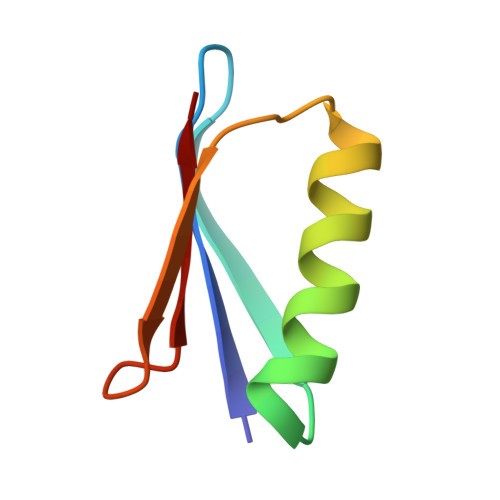
    Computational design and in vitro evolution are major strategies for stabilizing proteins. For the four critical positions 16, 18, 25, and 29 of the B domain of the streptococcal protein G (Gbeta1), they identified the same optimal residues at positions 16 and 25, but not at 18 and 29. Here we analyzed the energetic contributions of the residues from these two approaches by single and double mutant analyses and determined crystal structures for a variant from the calculation (I16/L18/E25/K29) and from the selection (I16/I18/E25/F29). The structural analysis explains the observed differences in stabilization. Residues 16, 18, and 29 line an invagination, which results from a packing defect between the helix and the beta-sheet of Gbeta1. In all stabilized variants, residues with larger side-chains occur at these positions and packing is improved. In the selected variant, packing is better optimized than in the computed variant. Such differences in side-chain packing strongly affect stability but are difficult to evaluate by computation.


  • Organizational Affiliation

    Laboratorium für Biochemie und Bayreuther Zentrum für Molekulare Biowissenschaften, Universität Bayreuth, D-95440 Bayreuth, Germany.


Macromolecules
Find similar proteins by:  (by identity cutoff)  |  3D Structure
Entity ID: 1
MoleculeChains Sequence LengthOrganismDetailsImage
Immunoglobulin G-binding protein G56Streptococcus sp. G148Mutation(s): 8 
Gene Names: spg
UniProt
Find proteins for P19909 (Streptococcus sp. group G)
Explore P19909 
Go to UniProtKB:  P19909
Entity Groups  
Sequence Clusters30% Identity50% Identity70% Identity90% Identity95% Identity100% Identity
UniProt GroupP19909
Sequence Annotations
Expand
  • Reference Sequence
Experimental Data & Validation

Experimental Data

  • Method: X-RAY DIFFRACTION
  • Resolution: 1.70 Å
  • R-Value Free: 0.229 
  • R-Value Work: 0.205 
  • R-Value Observed: 0.206 
  • Space Group: H 3 2
Unit Cell:
Length ( Å )Angle ( ˚ )
a = 48.412α = 90
b = 48.412β = 90
c = 107.332γ = 120
Software Package:
Software NamePurpose
XSCALEdata scaling
AMoREphasing
REFMACrefinement
PDB_EXTRACTdata extraction
MAR345dtbdata collection
XDSdata reduction

Structure Validation

View Full Validation Report


Entry History 

Deposition Data

Revision History  (Full details and data files)

  • Version 1.0: 2008-01-08
    Type: Initial release
  • Version 1.1: 2011-07-13
    Changes: Source and taxonomy, Version format compliance
  • Version 1.2: 2021-10-20
    Changes: Database references
  • Version 1.3: 2023-08-30
    Changes: Data collection, Refinement description