Solution structure of human secretory component and implications for biological function.
Bonner, A., Perrier, C., Corthesy, B., Perkins, S.J.(2007) J Biol Chem 282: 16969-16980
- PubMed: 17428798
- DOI: https://doi.org/10.1074/jbc.M701281200
- Primary Citation of Related Structures:
2OCW - PubMed Abstract:
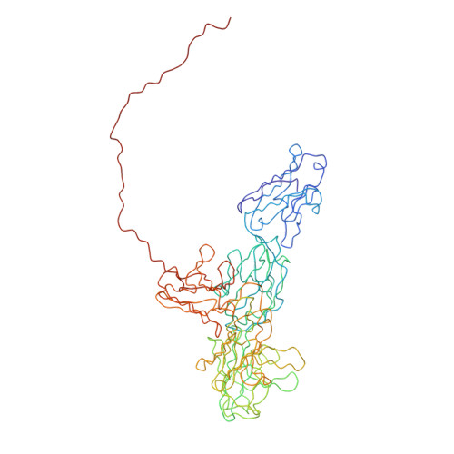
Secretory component (SC) in association with polymeric IgA (pIgA) forms secretory IgA, the major antibody active at mucosal surfaces. SC also exists in the free form, with innate-like neutralizing properties against pathogens. Free SC consists of five glycosylated variable (V)-type Ig domains (D1-D5), whose structure was determined by x-ray and neutron scattering, ultracentrifugation, and modeling. With a radius of gyration of 3.53-3.63 nm, a length of 12.5 nm, and a sedimentation coefficient of 4.0 S, SC possesses an unexpected compact structure. Constrained scattering modeling based on up to 13,000 trial models shows that SC adopts a J-shaped structure in which D4 and D5 are folded back against D2 and D3. The seven glycosylation sites are located on one side of SC, leaving known IgA-binding motifs free to interact with pIgA. This work represents the first analysis of the three-dimensional structure of full-length free SC and paves the way to a better understanding of the association between SC and its potential ligands, i.e. pIgA and pathogenic-associated motifs.
Organizational Affiliation:
Department of Biochemistry and Molecular Biology, University College London, Darwin Building, Gower Street, London WC1E 6BT, United Kingdom.