2O9U

Monellin (MNEI) at 1.15 resolution


Experimental Data Snapshot

  • Method: X-RAY DIFFRACTION
  • Resolution: 1.15 Å
  • R-Value Free: 0.165 
  • R-Value Work: 0.131 
  • R-Value Observed: 0.133 

wwPDB Validation   3D Report Full Report


This is version 1.4 of the entry. See complete history


Literature

Monellin (MNEI) at 1.15 A resolution

Hobbs, J.R.Munger, S.D.Conn, G.L.

(2007) Acta Crystallogr Sect F Struct Biol Cryst Commun 63: 162-167

  • DOI: https://doi.org/10.1107/S1744309107005271
  • Primary Citation of Related Structures:  
    2O9U

  • PubMed Abstract: 

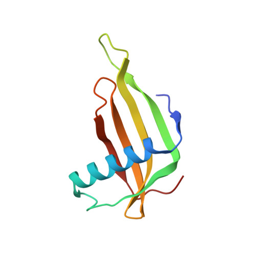
    The X-ray crystal structure of a single-chain monellin protein (MNEI) has been determined at 1.15 A resolution. The model was refined to convergence employing anisotropic displacement parameters and riding H atoms to produce a final model with R(work) and R(free) values of 0.132 and 0.162, respectively. The crystal contains a single MNEI protein in the asymmetric unit and unusually lacks the dimer interface observed in all previous crystal structures of monellin and its single-chain derivatives. The high resolution allowed a more detailed view of MNEI than previously possible, with 38 of the 96 residues modelled with alternative side-chain conformations, including four core residues Thr12, Cys41, Leu62 and Ile75. Four stably bound negative ions were also located, providing new insight into potential electrostatic interactions of MNEI with the largely negatively charged surface of the sweet taste receptor T1R2-T1R3.


  • Organizational Affiliation

    Manchester Interdisciplinary Biocentre, Faculty of Life Sciences, University of Manchester, Manchester M60 1QD, England.


Macromolecules
Find similar proteins by:  (by identity cutoff)  |  3D Structure
Entity ID: 1
MoleculeChains Sequence LengthOrganismDetailsImage
Monellin chain B and Monellin chain AA [auth X]97Dioscoreophyllum cumminsiiMutation(s): 0 
UniProt
Find proteins for P02882 (Dioscoreophyllum cumminsii)
Explore P02882 
Go to UniProtKB:  P02882
Entity Groups  
Sequence Clusters30% Identity50% Identity70% Identity90% Identity95% Identity100% Identity
UniProt GroupP02882
Sequence Annotations
Expand
  • Reference Sequence
Experimental Data & Validation

Experimental Data

  • Method: X-RAY DIFFRACTION
  • Resolution: 1.15 Å
  • R-Value Free: 0.165 
  • R-Value Work: 0.131 
  • R-Value Observed: 0.133 
  • Space Group: P 1 21 1
Unit Cell:
Length ( Å )Angle ( ˚ )
a = 27.103α = 90
b = 66.316β = 111.6
c = 27.154γ = 90
Software Package:
Software NamePurpose
REFMACrefinement
PDB_EXTRACTdata extraction
ADSCdata collection
MOSFLMdata reduction
CCP4data scaling
MOLREPphasing

Structure Validation

View Full Validation Report


Entry History 

Deposition Data

Revision History  (Full details and data files)

  • Version 1.0: 2007-02-20
    Type: Initial release
  • Version 1.1: 2008-05-01
    Changes: Version format compliance
  • Version 1.2: 2011-07-13
    Changes: Version format compliance
  • Version 1.3: 2017-08-16
    Changes: Advisory, Refinement description, Source and taxonomy
  • Version 1.4: 2023-10-25
    Changes: Advisory, Data collection, Database references, Derived calculations, Refinement description