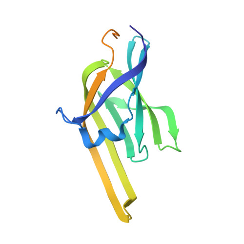
2O6P

Crystal Structure of the heme-IsdC complex


Experimental Data Snapshot

  • Method: X-RAY DIFFRACTION
  • Resolution: 1.50 Å
  • R-Value Free: 0.222 
  • R-Value Work: 0.193 
  • R-Value Observed: 0.195 

wwPDB Validation   3D Report Full Report


Ligand Structure Quality Assessment 


This is version 1.4 of the entry. See complete history


Literature

Crystal structure of the heme-IsdC complex, the central conduit of the Isd iron/heme uptake system in Staphylococcus aureus.

Sharp, K.H.Schneider, S.Cockayne, A.Paoli, M.

(2007) J Biol Chem 282: 10625-10631

  • DOI: https://doi.org/10.1074/jbc.M700234200
  • Primary Citation of Related Structures:  
    2O6P

  • PubMed Abstract: 

    Pathogens such as Staphylococcus aureus require iron to survive and have evolved specialized proteins to steal heme from their host. IsdC is the central conduit of the Isd (iron-regulated surface determinant) multicomponent heme uptake machinery; staphylococcal cell-surface proteins such as IsdA, IsdB, and IsdH are thought to funnel their molecular cargo to IsdC, which then mediates the transfer of the iron-containing nutrient to the membrane translocation system IsdDEF. The structure of the heme-IsdC complex reveals a novel heme site within an immunoglobulin-like domain and sheds light on its binding mechanism. The folding topology is reminiscent of the architecture of cytochrome f, cellobiose dehydrogenase, and ethylbenzene dehydrogenase; in these three proteins, the heme is bound in an equivalent position, but interestingly, IsdC features a distinct binding pocket with the ligand located next to the hydrophobic core of the beta-sandwich. The iron is coordinated with a tyrosine surrounded by several non-polar side chains that cluster into a tightly packed proximal side. On the other hand, the distal side is relatively exposed with a short helical peptide segment that acts as a lip clasping onto almost half of the porphyrin plane. This structural feature is argued to play a role in the mechanism of binding and release by switching to an open conformation and thus loosening the interactions holding the heme. The structure of the heme-IsdC complex provides a template for the understanding of other proteins, such as IsdA, IsdB, and IsdH, that contain the same heme-binding module as IsdC, known as the NEAT (near transporter) domain.


  • Organizational Affiliation

    School of Pharmacy, Centre for Biomolecular Sciences, University of Nottingham, University Park, Nottingham NG7 2RD, United Kingdom.


Macromolecules
Find similar proteins by:  (by identity cutoff)  |  3D Structure
Entity ID: 1
MoleculeChains Sequence LengthOrganismDetailsImage
Iron-regulated surface determinant protein C
A, B
161Staphylococcus aureus subsp. aureus N315Mutation(s): 0 
UniProt
Find proteins for Q8KQR1 (Staphylococcus aureus (strain NCTC 8325 / PS 47))
Explore Q8KQR1 
Go to UniProtKB:  Q8KQR1
Entity Groups  
Sequence Clusters30% Identity50% Identity70% Identity90% Identity95% Identity100% Identity
UniProt GroupQ8KQR1
Sequence Annotations
Expand
  • Reference Sequence
Small Molecules
Ligands 3 Unique
IDChains Name / Formula / InChI Key2D Diagram3D Interactions
HEM
Query on HEM

Download Ideal Coordinates CCD File 
F [auth A],
L [auth B]
PROTOPORPHYRIN IX CONTAINING FE
C34 H32 Fe N4 O4
KABFMIBPWCXCRK-RGGAHWMASA-L
ZN
Query on ZN

Download Ideal Coordinates CCD File 
C [auth A]
D [auth A]
G [auth B]
H [auth B]
I [auth B]
C [auth A],
D [auth A],
G [auth B],
H [auth B],
I [auth B],
J [auth B]
ZINC ION
Zn
PTFCDOFLOPIGGS-UHFFFAOYSA-N
CL
Query on CL

Download Ideal Coordinates CCD File 
E [auth A],
K [auth B]
CHLORIDE ION
Cl
VEXZGXHMUGYJMC-UHFFFAOYSA-M
Experimental Data & Validation

Experimental Data

  • Method: X-RAY DIFFRACTION
  • Resolution: 1.50 Å
  • R-Value Free: 0.222 
  • R-Value Work: 0.193 
  • R-Value Observed: 0.195 
  • Space Group: P 21 21 21
Unit Cell:
Length ( Å )Angle ( ˚ )
a = 47.489α = 90
b = 70.441β = 90
c = 88.685γ = 90
Software Package:
Software NamePurpose
REFMACrefinement
MOSFLMdata reduction
CCP4data scaling
SHARPphasing

Structure Validation

View Full Validation Report


Ligand Structure Quality Assessment 


Entry History 

Deposition Data

Revision History  (Full details and data files)

  • Version 1.0: 2007-02-06
    Type: Initial release
  • Version 1.1: 2008-05-01
    Changes: Version format compliance
  • Version 1.2: 2011-07-13
    Changes: Derived calculations, Source and taxonomy, Version format compliance
  • Version 1.3: 2017-12-13
    Changes: Advisory, Database references
  • Version 1.4: 2023-12-27
    Changes: Advisory, Data collection, Database references, Derived calculations