2O39

Human Adenovirus type 11 knob in complex with domains SCR1 and SCR2 of CD46 (membrane cofactor protein, MCP)


Experimental Data Snapshot

  • Method: X-RAY DIFFRACTION
  • Resolution: 2.85 Å
  • R-Value Free: 0.273 
  • R-Value Work: 0.238 
  • R-Value Observed: 0.238 

wwPDB Validation   3D Report Full Report


This is version 2.2 of the entry. See complete history


Literature

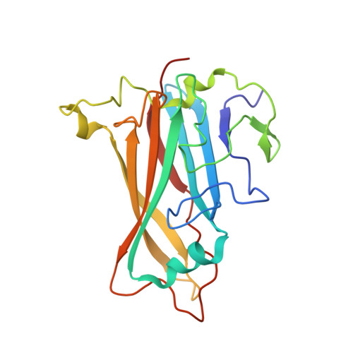
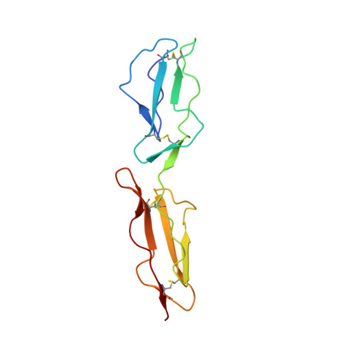
Adenovirus type 11 binding alters the conformation of its receptor CD46.

Persson, B.D.Reiter, D.M.Marttila, M.Mei, Y.F.Casasnovas, J.M.Arnberg, N.Stehle, T.

(2007) Nat Struct Mol Biol 14: 164-166

  • DOI: https://doi.org/10.1038/nsmb1190
  • Primary Citation of Related Structures:  
    2O39

  • PubMed Abstract: 

    Adenoviruses (Ads) are important human pathogens and valuable gene delivery vehicles. We report here the crystal structure of the species B Ad11 knob complexed with the Ad11-binding region of its receptor CD46. The conformation of bound CD46 differs profoundly from its unbound state, with the bent surface structure straightened into an elongated rod. This mechanism of interaction is likely to be conserved among many pathogens that target CD46 or related molecules.


  • Organizational Affiliation

    Interfaculty Institute for Biochemistry, University of Tübingen, D-72076 Tübingen, Germany.


Macromolecules
Find similar proteins by:  (by identity cutoff)  |  3D Structure
Entity ID: 1
MoleculeChains Sequence LengthOrganismDetailsImage
Fiber protein
A, B
197Human adenovirus 11pMutation(s): 0 
Gene Names: PIV
UniProt
Find proteins for P35774 (Human adenovirus B serotype 11 (strain Slobiski))
Explore P35774 
Go to UniProtKB:  P35774
Entity Groups  
Sequence Clusters30% Identity50% Identity70% Identity90% Identity95% Identity100% Identity
UniProt GroupP35774
Sequence Annotations
Expand
  • Reference Sequence
Find similar proteins by:  (by identity cutoff)  |  3D Structure
Entity ID: 2
MoleculeChains Sequence LengthOrganismDetailsImage
Membrane cofactor protein
C, D
126Homo sapiensMutation(s): 0 
Gene Names: CD46MCP
UniProt & NIH Common Fund Data Resources
Find proteins for P15529 (Homo sapiens)
Explore P15529 
Go to UniProtKB:  P15529
PHAROS:  P15529
GTEx:  ENSG00000117335 
Entity Groups  
Sequence Clusters30% Identity50% Identity70% Identity90% Identity95% Identity100% Identity
UniProt GroupP15529
Sequence Annotations
Expand
  • Reference Sequence
Oligosaccharides

Help

Entity ID: 3
MoleculeChains Length2D Diagram Glycosylation3D Interactions
beta-D-mannopyranose-(1-4)-2-acetamido-2-deoxy-beta-D-glucopyranose-(1-4)-2-acetamido-2-deoxy-beta-D-glucopyranose
E, F
3N-Glycosylation
Glycosylation Resources
GlyTouCan:  G15407YE
GlyCosmos:  G15407YE
GlyGen:  G15407YE
Experimental Data & Validation

Experimental Data

  • Method: X-RAY DIFFRACTION
  • Resolution: 2.85 Å
  • R-Value Free: 0.273 
  • R-Value Work: 0.238 
  • R-Value Observed: 0.238 
  • Space Group: P 3
Unit Cell:
Length ( Å )Angle ( ˚ )
a = 106.14α = 90
b = 106.14β = 90
c = 68.32γ = 120
Software Package:
Software NamePurpose
HKL-2000data collection
AMoREphasing
CNSrefinement
DENZOdata reduction
SCALEPACKdata scaling

Structure Validation

View Full Validation Report


Entry History 

Deposition Data

Revision History  (Full details and data files)

  • Version 1.0: 2007-01-09
    Type: Initial release
  • Version 1.1: 2008-05-01
    Changes: Version format compliance
  • Version 1.2: 2011-07-13
    Changes: Non-polymer description, Version format compliance
  • Version 2.0: 2020-07-29
    Type: Remediation
    Reason: Carbohydrate remediation
    Changes: Atomic model, Data collection, Derived calculations, Structure summary
  • Version 2.1: 2023-12-27
    Changes: Data collection, Database references, Structure summary
  • Version 2.2: 2024-04-03
    Changes: Refinement description