2NN4

Crystal structure of Bacillus subtilis yqgQ, Pfam DUF910


Experimental Data Snapshot

  • Method: X-RAY DIFFRACTION
  • Resolution: 2.10 Å
  • R-Value Free: 0.280 
  • R-Value Work: 0.247 
  • R-Value Observed: 0.247 

wwPDB Validation   3D Report Full Report


This is version 1.5 of the entry. See complete history


Literature

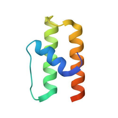
Structure of YqgQ protein from Bacillus subtilis, a conserved hypothetical protein.

Lakshminarasimhan, D.Eswaramoorthy, S.Burley, S.K.Swaminathan, S.

(2010) Acta Crystallogr Sect F Struct Biol Cryst Commun 66: 8-11

  • DOI: https://doi.org/10.1107/S1744309109047009
  • Primary Citation of Related Structures:  
    2NN4

  • PubMed Abstract: 

    The crystal structure of the hypothetical protein YqgQ from Bacillus subtilis has been determined to 2.1 A resolution. The crystals belonged to space group P2(1), with unit-cell parameters a = 51.85, b = 41.25, c = 55.18 A, beta = 113.4 degrees , and contained three protein molecules in the asymmetric unit. The structure was determined by the single-wavelength anomalous dispersion method using selenium-labeled protein and was refined to a final R factor of 24.7% (R(free) = 28.0%). The protein molecule mainly comprises a three-helical bundle. Its putative function is inferred to be single-stranded nucleic acid binding based on sequence and structural homology.


  • Organizational Affiliation

    Biology Department, Brookhaven National Laboratory, Upton, New York 11973, USA.


Macromolecules
Find similar proteins by:  (by identity cutoff)  |  3D Structure
Entity ID: 1
MoleculeChains Sequence LengthOrganismDetailsImage
Hypothetical protein yqgQ
A, B, C
72Bacillus subtilisMutation(s): 0 
Gene Names: yqgQ
UniProt
Find proteins for P54494 (Bacillus subtilis (strain 168))
Explore P54494 
Go to UniProtKB:  P54494
Entity Groups  
Sequence Clusters30% Identity50% Identity70% Identity90% Identity95% Identity100% Identity
UniProt GroupP54494
Sequence Annotations
Expand
  • Reference Sequence
Experimental Data & Validation

Experimental Data

  • Method: X-RAY DIFFRACTION
  • Resolution: 2.10 Å
  • R-Value Free: 0.280 
  • R-Value Work: 0.247 
  • R-Value Observed: 0.247 
  • Space Group: P 1 21 1
Unit Cell:
Length ( Å )Angle ( ˚ )
a = 51.85α = 90
b = 41.25β = 113.39
c = 55.18γ = 90
Software Package:
Software NamePurpose
CNSrefinement
CBASSdata collection
HKL-2000data reduction
HKL-2000data scaling
SOLVEphasing
SHARPphasing

Structure Validation

View Full Validation Report


Entry History 

Revision History  (Full details and data files)

  • Version 1.0: 2006-10-31
    Type: Initial release
  • Version 1.1: 2008-05-01
    Changes: Version format compliance
  • Version 1.2: 2011-07-13
    Changes: Version format compliance
  • Version 1.3: 2012-10-24
    Changes: Database references
  • Version 1.4: 2021-02-03
    Changes: Database references, Structure summary
  • Version 1.5: 2023-12-27
    Changes: Data collection, Database references