Analysis of the Structural and Molecular Basis of Voltage-sensitive Sodium Channel Inhibition by the Spider Toxin Huwentoxin-IV ( mu-TRTX-Hh2a).
Minassian, N.A., Gibbs, A., Shih, A.Y., Liu, Y., Neff, R.A., Sutton, S.W., Mirzadegan, T., Connor, J., Fellows, R., Husovsky, M., Nelson, S., Hunter, M.J., Flinspach, M., Wickenden, A.D.(2013) J Biol Chem 288: 22707-22720
- PubMed: 23760503
- DOI: https://doi.org/10.1074/jbc.M113.461392
- Primary Citation of Related Structures:
2M4X, 2M4Z, 2M50 - PubMed Abstract:
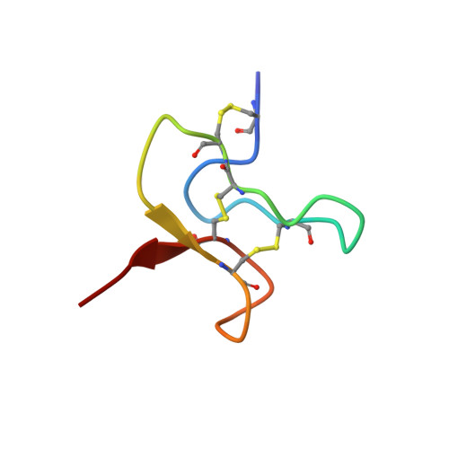
Voltage-gated sodium channels (VGSCs) are essential to the normal function of the vertebrate nervous system. Aberrant function of VGSCs underlies a variety of disorders, including epilepsy, arrhythmia, and pain. A large number of animal toxins target these ion channels and may have significant therapeutic potential. Most of these toxins, however, have not been characterized in detail. Here, by combining patch clamp electrophysiology and radioligand binding studies with peptide mutagenesis, NMR structure determination, and molecular modeling, we have revealed key molecular determinants of the interaction between the tarantula toxin huwentoxin-IV and two VGSC isoforms, Nav1.7 and Nav1.2. Nine huwentoxin-IV residues (F6A, P11A, D14A, L22A, S25A, W30A, K32A, Y33A, and I35A) were important for block of Nav1.7 and Nav1.2. Importantly, molecular dynamics simulations and NMR studies indicated that folding was normal for several key mutants, suggesting that these amino acids probably make specific interactions with sodium channel residues. Additionally, we identified several amino acids (F6A, K18A, R26A, and K27A) that are involved in isoform-specific VGSC interactions. Our structural and functional data were used to model the docking of huwentoxin-IV into the domain II voltage sensor of Nav1.7. The model predicts that a hydrophobic patch composed of Trp-30 and Phe-6, along with the basic Lys-32 residue, docks into a groove formed by the Nav1.7 S1-S2 and S3-S4 loops. These results provide new insight into the structural and molecular basis of sodium channel block by huwentoxin-IV and may provide a basis for the rational design of toxin-based peptides with improved VGSC potency and/or selectivity.
Organizational Affiliation:
Department of Neuroscience Discovery, Janssen Research & Development, LLC, San Diego, California 92121, USA.