Structure determination of a membrane protein in proteoliposomes.
Das, B.B., Nothnagel, H.J., Lu, G.J., Son, W.S., Tian, Y., Marassi, F.M., Opella, S.J.(2012) J Am Chem Soc 134: 2047-2056
- PubMed: 22217388
- DOI: https://doi.org/10.1021/ja209464f
- Primary Citation of Related Structures:
2LJ2 - PubMed Abstract:
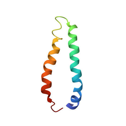
An NMR method for determining the three-dimensional structures of membrane proteins in proteoliposomes is demonstrated by determining the structure of MerFt, the 60-residue helix-loop-helix integral membrane core of the 81-residue mercury transporter MerF. The method merges elements of oriented sample (OS) solid-state NMR and magic angle spinning (MAS) solid-state NMR techniques to measure orientation restraints relative to a single external axis (the bilayer normal) from individual residues in a uniformly (13)C/(15)N labeled protein in unoriented liquid crystalline phospholipid bilayers. The method relies on the fast (>10(5) Hz) rotational diffusion of membrane proteins in bilayers to average the static chemical shift anisotropy and heteronuclear dipole-dipole coupling powder patterns to axially symmetric powder patterns with reduced frequency spans. The frequency associated with the parallel edge of such motionally averaged powder patterns is exactly the same as that measured from the single line resonance in the spectrum of a stationary sample that is macroscopically aligned parallel to the direction of the applied magnetic field. All data are collected on unoriented samples undergoing MAS. Averaging of the homonuclear (13)C/(13)C dipolar couplings, by MAS of the sample, enables the use of uniformly (13)C/(15)N labeled proteins, which provides enhanced sensitivity through direct (13)C detection as well as the use of multidimensional MAS solid-state NMR methods for resolving and assigning resonances. The unique feature of this method is the measurement of orientation restraints that enable the protein structure and orientation to be determined in unoriented proteoliposomes.
Organizational Affiliation:
Department of Chemistry and Biochemistry, University of California, San Diego, La Jolla, California 92093-0307, USA.