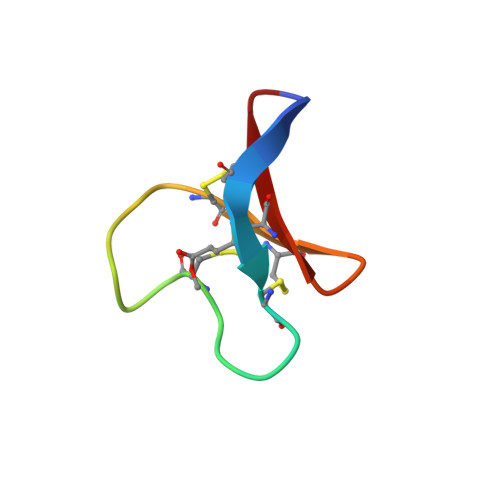
The conserved glu in the cyclotide cycloviolacin O2 has a key structural role.
Goransson, U., Herrmann, A., Burman, R., Haugaard-Jonsson, L.M., Rosengren, K.J.(2009) Chembiochem 10: 2354-2360
- PubMed: 19735083
- DOI: https://doi.org/10.1002/cbic.200900342
- Primary Citation of Related Structures:
2KNM, 2KNN - PubMed Abstract:
Cyclotides are a large family of plant peptides that are characterised by a head-to-tail circular backbone and three disulfide bonds that are arranged in a cystine knot. This unique structural feature, which is referred to as a cyclic cystine knot, gives the cyclotides remarkable stability against chemical and biological degradation. In addition to their natural function as insecticides for plant defence, the cyclotides have a range of bioactivities with pharmaceutical relevance, including cytotoxicity against cancer cell lines. A glutamic acid residue, aside from the invariable disulfide array, is the most conserved feature throughout the cyclotide family, and it has recently been shown to be crucial for biological activity. Here we have used solution-state NMR spectroscopy to determine the three-dimensional structures of the potent cytotoxic cyclotide cycloviolacin O2, and an inactive analogue in which this conserved glutamic acid has been methylated. The structures of the peptides show that the glutamic acid has a key structural role in coordinating a set of hydrogen bonds in native cycloviolacin O2; this interaction is disrupted in the methylated analogue. The proposed mechanism of action of cyclotides is membrane disruption and these results suggest that the glutamic acid is linked to cyclotide function by stabilising the structure to allow efficient aggregation in membranes, rather than in a direct interaction with a target receptor.
Organizational Affiliation:
Division of Pharmacognosy, Department of Medicinal Chemistry, Biomedical Centre, Uppsala University, 751 23 Uppsala, Sweden.