Solution structure and interaction of cupiennin 1a, a spider venom peptide, with phospholipid bilayers
Pukala, T.L., Boland, M.P., Gehman, J.D., Kuhn-Nentwig, L., Separovic, F., Bowie, J.H.(2007) Biochemistry 46: 3576-3585
- PubMed: 17319697
- DOI: https://doi.org/10.1021/bi062306+
- Primary Citation of Related Structures:
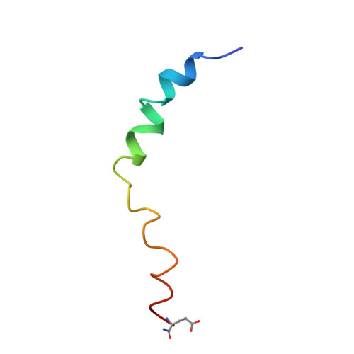
2K38 - PubMed Abstract:
The solution structure of cupiennin 1a, a 35 residue, basic antibacterial peptide isolated from the venom of the spider Cupiennius salei, has been determined by nuclear magnetic resonance (NMR) spectroscopy. The peptide was found to adopt a helix-hinge-helix structure in a membrane mimicking solvent. The hinge may play a role in allowing the amphipathic N-terminal helix and polar C-terminal helix to orient independently upon membrane binding, in order to achieve maximal antibacterial efficacy. Solid-state 31P and 2H NMR was used to further study the effects of cupiennin 1a on the dynamic properties of lipid membranes, using zwitterionic chain deuterated dimyristoylphosphatidylcholine (d54-DMPC) and anionic dimyristoylphosphatidylglycerol (DMPG) multilamellar vesicles. In d54-DMPC alone, cupiennin 1a caused a decrease in the 31P chemical shift anisotropy, indicating some interaction with the lipid head groups, and a decrease in order over the entire acyl chain. In contrast, for the mixed (d54-DMPC/DMPG) lipid system cupiennin 1a appeared to induce lateral separation of the two lipids as evidenced by the 31P spectra, in which the peptide preferentially interacted with DMPG. Little effect was observed on the deuterated acyl chain order parameters in the d54-DMPC/DMPG model membranes. Furthermore, 31P NMR relaxation measurements confirmed a differential effect on the lipid motions depending upon the membrane composition. Therefore, subtle differences are likely in the mechanism by which cupiennin 1a causes membrane lysis in either prokaryotic or eukaryotic cells, and may explain the specific spectrum of activity.
Organizational Affiliation:
Department of Chemistry, University of Adelaide, Adelaide, South Australia, 5005, Australia.