Solution structure of the yeast URN1 splicing factor FF domain: Comparative analysis of charge distributions in FF domain structures-FFs and SURPs, two domains with a similar fold.
Bonet, R., Ramirez-Espain, X., Macias, M.J.(2008) Proteins 73: 1001-1009
- PubMed: 18536009
- DOI: https://doi.org/10.1002/prot.22127
- Primary Citation of Related Structures:
2JUC - PubMed Abstract:
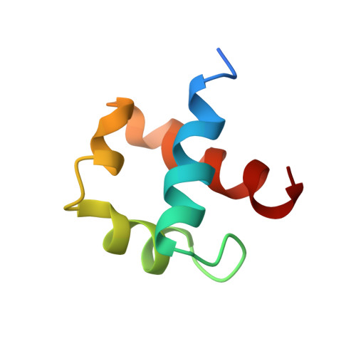
FF domains are present in three protein families: the splicing factors formin binding protein 11 (FBP11), Prp40, and URN1, the transcription factor CA150, and the p190RhoGTPase-related proteins. This simplicity in distribution, however, is contrasted by the difficulty in defining their biological role. At best, the group of ligand FF domains can bind to form a motley crew with binding reports pointing also to negative/aromatic sequences, the tetratricopeptide repeat, the transcription factor TFII-I and even to RNA. To expand our knowledge on the FF domain, we selected the FF domain present in the URN1 yeast splicing factor as the subject for structural studies. The URN1 protein is one of the two known proteins containing only one FF domain, making it the most simplified representative of FF domain-containing splicing factors. The solution structure reveals that the domain adopts the classical FF fold, with a distinctive negatively charged patch on its surface. All available FF structures have a well-conserved fold but variable electrostatic patches on their surfaces. These patches are unconserved, even for domains with similar pK(a)s. To investigate potential binding sites in FF domains, we performed structural comparisons to other proteins with similar folds. In addition to the structures detected by SCOP, we included SURP domains, which also adopt the alpha1-alpha2-3(10)-alpha3 architecture. We observed that the main difference between all these structures resides in the orientation of the second helix. Remarkably, in DEK, SURP, and Prp40FF1 structures (the exception is the FBP11FF1 domain), the second helix participates in ligand recognition. Furthermore, SURP and Prp40FF1 binding sites also include the 3(10) helix, which forms a partially exposed hydrophobic cavity. This cavity is also present in at least CA150FF1 and FF2 structures. Thus, as with WW domains, the FF fold seems to have developed binding-site variations to accommodate an abundant and variable set of ligands.
Organizational Affiliation:
The Institute for Research in Biomedicine, Barcelona (Protein NMR Group), Barcelona Science Park, Baldiri i Reixac, 10-12 08028 Barcelona, Spain.