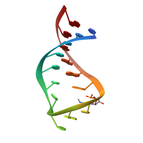
Anticodon domain modifications contribute order to tRNA for ribosome-mediated codon binding.
Vendeix, F.A., Dziergowska, A., Gustilo, E.M., Graham, W.D., Sproat, B., Malkiewicz, A., Agris, P.F.(2008) Biochemistry 47: 6117-6129
- PubMed: 18473483
- DOI: https://doi.org/10.1021/bi702356j
- Primary Citation of Related Structures:
2JR4, 2JRG - PubMed Abstract:
The accuracy and efficiency with which tRNA decodes genomic information into proteins require posttranscriptional modifications in or adjacent to the anticodon. The modification uridine-5-oxyacetic acid (cmo (5)U 34) is found at wobble position 34 in a single isoaccepting tRNA species for six amino acids, alanine, leucine, proline, serine, threonine, and valine, each having 4-fold degenerate codons. cmo (5)U 34 makes possible the decoding of 24 codons by just six tRNAs. The contributions of this important modification to the structures and codon binding affinities of the unmodified and fully modified anticodon stem and loop domains of tRNA (Val3) UAC (ASL (Val3) UAC) were elucidated. The stems of the unmodified ASL (Val3) UAC and that with cmo (5)U 34 and N (6)-methyladenosine, m (6)A 37, adopted an A-form RNA conformation (rmsd approximately 0.6 A) as determined with NMR spectroscopy and torsion-angle molecular dynamics. However, the UV hyperchromicity, circular dichroism ellipticity, and structural analyses indicated that the anticodon modifications enhanced order in the loop. ASL (Val3) UAC-cmo (5)U 34;m (6)A 37 exhibited high affinities for its cognate and wobble codons GUA and GUG, and for GUU in the A-site of the programmed 30S ribosomal subunit, whereas the unmodified ASL (Val3) UAC bound less strongly to GUA and not at all to GUG and GUU. Together with recent crystal structures of ASL (Val3) UAC-cmo (5)U 34;m (6)A 37 bound to all four of the valine codons in the A-site of the ribosome's 30S subunit, these results clearly demonstrate that the xo (5)U 34-type modifications order the anticodon loop prior to A-site codon binding for an expanded codon reading, possibly reducing an entropic energy barrier to codon binding.
Organizational Affiliation:
Department of Molecular and Structural Biochemistry, North Carolina State University, 128 Polk Hall, Raleigh, North Carolina 27695-7622, USA.