2JJV

Structure of human signal regulatory protein (sirp) beta(2)


Experimental Data Snapshot

  • Method: X-RAY DIFFRACTION
  • Resolution: 1.80 Å
  • R-Value Free: 0.213 
  • R-Value Work: 0.172 
  • R-Value Observed: 0.174 

wwPDB Validation   3D Report Full Report


This is version 1.4 of the entry. See complete history


Literature

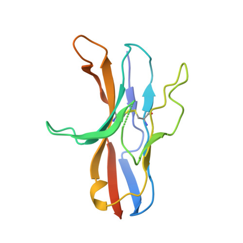
Paired Receptor Specificity Explained by Structures of Signal Regulatory Proteins Alone and Complexed with Cd47.

Hatherley, D.Graham, S.C.Turner, J.Harlos, K.Stuart, D.I.Barclay, A.N.

(2008) Mol Cell 31: 266

  • DOI: https://doi.org/10.1016/j.molcel.2008.05.026
  • Primary Citation of Related Structures:  
    2JJS, 2JJT, 2JJU, 2JJV, 2JJW, 2VSC

  • PubMed Abstract: 

    CD47 is a widely distributed cell-surface protein that acts a marker of self through interactions of myeloid and neural cells. We describe the high-resolution X-ray crystallographic structures of the immunoglobulin superfamily domain of CD47 alone and in complex with the N-terminal ligand-binding domain of signal regulatory protein alpha (SIRPalpha). The unusual and convoluted interacting face of CD47, comprising the N terminus and loops at the end of the domain, intercalates with the corresponding regions in SIRPalpha. We have also determined structures of the N-terminal domains of SIRPbeta, SIRPbeta(2), and SIRPgamma; proteins that are closely related to SIRPalpha but bind CD47 with negligible or reduced affinity. These results explain the specificity of CD47 for the SIRP family of paired receptors in atomic detail. Analysis of SIRPalpha polymorphisms suggests that these, as well as the activating SIRPs, may have evolved to counteract pathogen binding to the inhibitory SIRPalpha receptor.


  • Organizational Affiliation

    Sir William Dunn School of Pathology, University of Oxford, Oxford, OX1 3RE, UK.


Macromolecules
Find similar proteins by:  (by identity cutoff)  |  3D Structure
Entity ID: 1
MoleculeChains Sequence LengthOrganismDetailsImage
SIGNAL-REGULATORY PROTEIN BETA 1.
A, B
127Homo sapiensMutation(s): 0 
UniProt & NIH Common Fund Data Resources
Find proteins for Q5TFQ8 (Homo sapiens)
Explore Q5TFQ8 
Go to UniProtKB:  Q5TFQ8
PHAROS:  Q5TFQ8
GTEx:  ENSG00000101307 
Entity Groups  
Sequence Clusters30% Identity50% Identity70% Identity90% Identity95% Identity100% Identity
UniProt GroupQ5TFQ8
Sequence Annotations
Expand
  • Reference Sequence
Experimental Data & Validation

Experimental Data

  • Method: X-RAY DIFFRACTION
  • Resolution: 1.80 Å
  • R-Value Free: 0.213 
  • R-Value Work: 0.172 
  • R-Value Observed: 0.174 
  • Space Group: C 1 2 1
Unit Cell:
Length ( Å )Angle ( ˚ )
a = 114.979α = 90
b = 47.49β = 93.39
c = 40.044γ = 90
Software Package:
Software NamePurpose
REFMACrefinement
HKL-2000data reduction
HKL-2000data scaling
MOLREPphasing

Structure Validation

View Full Validation Report


Entry History 

Deposition Data

Revision History  (Full details and data files)

  • Version 1.0: 2008-08-05
    Type: Initial release
  • Version 1.1: 2011-05-07
    Changes: Version format compliance
  • Version 1.2: 2011-07-13
    Changes: Version format compliance
  • Version 1.3: 2019-04-03
    Changes: Data collection, Other, Source and taxonomy
  • Version 1.4: 2023-12-13
    Changes: Data collection, Database references, Other, Refinement description