2JFC

M144L mutant of Nitrite Reductase from Alcaligenes xylosoxidans in space group P212121


Experimental Data Snapshot

  • Method: X-RAY DIFFRACTION
  • Resolution: 2.40 Å
  • R-Value Free: 0.193 
  • R-Value Work: 0.171 
  • R-Value Observed: 0.172 

wwPDB Validation   3D Report Full Report


This is version 1.3 of the entry. See complete history


Literature

The Structure of the met144Leu Mutant of Copper Nitrite Reductase from Alcaligenes Xylosoxidans Provides the First Glimpse of a Protein-Protein Complex with Azurin II.

Paraskevopoulos, K.Hough, M.A.Sawers, R.G.Eady, R.R.Hasnain, S.S.

(2007) J Biol Inorg Chem 12: 789

  • DOI: https://doi.org/10.1007/s00775-007-0233-y
  • Primary Citation of Related Structures:  
    2JFC

  • PubMed Abstract: 

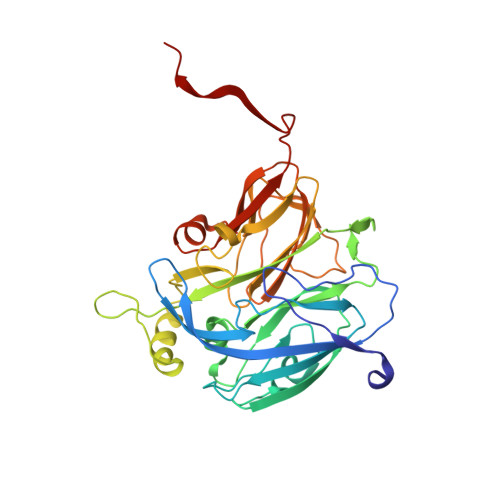
    Cu-containing nitrite reductases (NiRs) perform the reduction of nitrite to NO via an ordered mechanism in which the delivery of a proton and an electron to the catalytic type 2 Cu site is highly orchestrated. Electron transfer from a redox partner protein, azurin or pseudoazurin, to the type 1 Cu site is assumed to occur through the formation of a protein-protein complex. We report here a new crystal form in space group P2(1)2(1)2(1) of the Met144Leu mutant of NiR from Alcaligenes xylosoxidans (AxNiR), revealing a head-to-head packing motif involving residues around the hydrophobic patch of domain 1. Superposition of the structure of azurin II with that of domain 1 of one of the Met144Leu molecules provides the first glimpse of an azurin II-NiR protein-protein complex. Mutations of two of the residues of AxNiR, Trp138His (Barrett et al. in Biochemistry 43:16311-16319, 2004) and Met87Leu, highlighted in the AxNiR-azurin complex, results in substantially decreased activity when azurin is used as the electron donor instead of methyl viologen, providing direct evidence for the importance of this region for complex formation.


  • Organizational Affiliation

    Molecular Biophysics Group, STFC Daresbury Laboratory, Warrington, UK.


Macromolecules
Find similar proteins by:  (by identity cutoff)  |  3D Structure
Entity ID: 1
MoleculeChains Sequence LengthOrganismDetailsImage
DISSIMILATORY COPPER-CONTAINING NITRITE REDUCTASE
A, B, C, D, E
A, B, C, D, E, F
335Achromobacter xylosoxidansMutation(s): 1 
EC: 1.7.2.1
UniProt
Find proteins for O68601 (Alcaligenes xylosoxydans xylosoxydans)
Explore O68601 
Go to UniProtKB:  O68601
Entity Groups  
Sequence Clusters30% Identity50% Identity70% Identity90% Identity95% Identity100% Identity
UniProt GroupO68601
Sequence Annotations
Expand
  • Reference Sequence
Small Molecules
Ligands 2 Unique
IDChains Name / Formula / InChI Key2D Diagram3D Interactions
CU
Query on CU

Download Ideal Coordinates CCD File 
G [auth A]
H [auth A]
J [auth B]
K [auth B]
M [auth C]
G [auth A],
H [auth A],
J [auth B],
K [auth B],
M [auth C],
N [auth C],
P [auth D],
Q [auth D],
R [auth E],
S [auth E],
T [auth F],
U [auth F]
COPPER (II) ION
Cu
JPVYNHNXODAKFH-UHFFFAOYSA-N
CL
Query on CL

Download Ideal Coordinates CCD File 
I [auth A],
L [auth B],
O [auth C],
V [auth F]
CHLORIDE ION
Cl
VEXZGXHMUGYJMC-UHFFFAOYSA-M
Experimental Data & Validation

Experimental Data

  • Method: X-RAY DIFFRACTION
  • Resolution: 2.40 Å
  • R-Value Free: 0.193 
  • R-Value Work: 0.171 
  • R-Value Observed: 0.172 
  • Space Group: P 21 21 21
Unit Cell:
Length ( Å )Angle ( ˚ )
a = 171.958α = 90
b = 175.91β = 90
c = 181.056γ = 90
Software Package:
Software NamePurpose
REFMACrefinement
HKL-2000data reduction
HKL-2000data scaling
MOLREPphasing

Structure Validation

View Full Validation Report


Entry History 

Deposition Data

Revision History  (Full details and data files)

  • Version 1.0: 2007-05-29
    Type: Initial release
  • Version 1.1: 2011-05-08
    Changes: Version format compliance
  • Version 1.2: 2011-07-13
    Changes: Version format compliance
  • Version 1.3: 2023-12-13
    Changes: Data collection, Database references, Derived calculations, Other, Refinement description