2JAG

L1-intermediate of halorhodopsin T203V


Experimental Data Snapshot

  • Method: X-RAY DIFFRACTION
  • Resolution: 1.93 Å
  • R-Value Free: 0.261 
  • R-Value Work: 0.237 
  • R-Value Observed: 0.237 

wwPDB Validation   3D Report Full Report


This is version 1.4 of the entry. See complete history


Literature

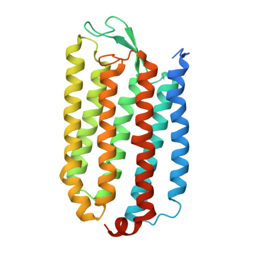
The crystal structure of the L1 intermediate of halorhodopsin at 1.9 angstroms resolution.

Gmelin, W.Zeth, K.Efremov, R.Heberle, J.Tittor, J.Oesterhelt, D.

(2007) Photochem Photobiol 83: 369-377

  • DOI: https://doi.org/10.1562/2006-06-23-RA-947
  • Primary Citation of Related Structures:  
    2JAF, 2JAG

  • PubMed Abstract: 

    The mutant T203V of the light driven chloride pump halorhodopsin from Halobacterium salinarum was crystallized and the X-ray structure was solved at 1.6 angstroms resolution. The T203V structure turned out to be nearly identical to the wild type protein with a root mean square deviation of 0.43 angstroms for the carbon alpha atoms of the protein backbone. Two chloride binding (CB) sites were demonstrated by a substitution of chloride with bromide and an analysis of anomalous difference Fourier maps. The CB1 site was found at the same position as in the wild type structure. In addition, a second chloride binding site CB2 was identified around Q105 due to higher resolution in the mutant crystal. As T203V showed a 10 times slower decay of its photocycle intermediate L, this intermediate could be trapped with an occupancy of 60% upon illumination at room temperature and subsequent cooling to 120 degrees K. Fourier transform infrared spectroscopy clearly identified the crystal to be trapped in the L1 intermediate state and the X-ray structure was solved to 1.9 angstroms resolution. In this intermediate, the chloride moved by 0.3 angstroms within binding site CB1 as indicated by peaks in difference Fourier density maps. The chloride in the second binding site CB2 remained unchanged. Thus, intraproteinous chloride translocation from the extracellular to the cytoplasmic part of the protein must occur in reaction steps following the L1 intermediate in the catalytic cycle of halorhodopsin.


  • Organizational Affiliation

    Max Planck Institute of Biochemistry, Department of Membrane Biochemistry, Martinsried, Germany.


Macromolecules
Find similar proteins by:  (by identity cutoff)  |  3D Structure
Entity ID: 1
MoleculeChains Sequence LengthOrganismDetailsImage
Halorhodopsin274Halobacterium salinarum R1Mutation(s): 2 
Gene Names: hopOE_1299R
Membrane Entity: Yes 
UniProt
Find proteins for B0R2U4 (Halobacterium salinarum (strain ATCC 29341 / DSM 671 / R1))
Explore B0R2U4 
Go to UniProtKB:  B0R2U4
Entity Groups  
Sequence Clusters30% Identity50% Identity70% Identity90% Identity95% Identity100% Identity
UniProt GroupB0R2U4
Sequence Annotations
Expand
  • Reference Sequence
Small Molecules
Ligands 4 Unique
IDChains Name / Formula / InChI Key2D Diagram3D Interactions
BOG
Query on BOG

Download Ideal Coordinates CCD File 
E [auth A]octyl beta-D-glucopyranoside
C14 H28 O6
HEGSGKPQLMEBJL-RKQHYHRCSA-N
RET
Query on RET

Download Ideal Coordinates CCD File 
F [auth A]RETINAL
C20 H28 O
NCYCYZXNIZJOKI-OVSJKPMPSA-N
PLM
Query on PLM

Download Ideal Coordinates CCD File 
D [auth A]PALMITIC ACID
C16 H32 O2
IPCSVZSSVZVIGE-UHFFFAOYSA-N
CL
Query on CL

Download Ideal Coordinates CCD File 
B [auth A],
C [auth A]
CHLORIDE ION
Cl
VEXZGXHMUGYJMC-UHFFFAOYSA-M
Experimental Data & Validation

Experimental Data

  • Method: X-RAY DIFFRACTION
  • Resolution: 1.93 Å
  • R-Value Free: 0.261 
  • R-Value Work: 0.237 
  • R-Value Observed: 0.237 
  • Space Group: H 3 2
Unit Cell:
Length ( Å )Angle ( ˚ )
a = 95.042α = 90
b = 95.042β = 90
c = 157.674γ = 120
Software Package:
Software NamePurpose
CNSrefinement
XDSdata reduction
AMoREphasing

Structure Validation

View Full Validation Report


Entry History 

Deposition Data

Revision History  (Full details and data files)

  • Version 1.0: 2006-12-14
    Type: Initial release
  • Version 1.1: 2011-12-28
    Changes: Database references, Derived calculations, Non-polymer description, Other, Refinement description
  • Version 1.2: 2018-11-21
    Changes: Data collection, Database references, Source and taxonomy, Structure summary
  • Version 1.3: 2020-07-29
    Type: Remediation
    Reason: Carbohydrate remediation
    Changes: Data collection, Derived calculations, Structure summary
  • Version 1.4: 2023-12-13
    Changes: Data collection, Database references, Refinement description, Structure summary