2J9A

blLAP in Complex with Microginin FR1


Experimental Data Snapshot

  • Method: X-RAY DIFFRACTION
  • Resolution: 1.73 Å
  • R-Value Free: 0.173 
  • R-Value Work: 0.145 
  • R-Value Observed: 0.146 

wwPDB Validation   3D Report Full Report


Ligand Structure Quality Assessment 


This is version 1.4 of the entry. See complete history


Literature

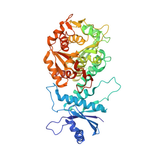
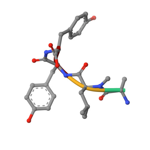
Binding Structure of the Leucine Aminopeptidase Inhibitor Microginin Fr1.

Kraft, M.Schleberger, C.Weckesser, J.Schulz, G.E.

(2006) FEBS Lett 580: 6943

  • DOI: https://doi.org/10.1016/j.febslet.2006.11.060
  • Primary Citation of Related Structures:  
    2J9A

  • PubMed Abstract: 

    Natural bioactive compounds are of general interest for pharmaceutical research because they may serve as leads in drug development campaigns. Among them, microginins are linear peptides known to inhibit various exopeptidases. The crystal structure of microginin FR1 from Microcystis sp. bound to bovine lens leucine aminopeptidase was established at 1.73 Angstrom resolution. The observed binding structure could be beneficial for the design of potent aminopeptidase inhibitors.


  • Organizational Affiliation

    Institut für Biologie II, Microbiologie, Albert-Ludwigs-Universität, Schänzlestr. 1, D-79104 Freiburg im Breisgau, Germany.


Macromolecules
Find similar proteins by:  (by identity cutoff)  |  3D Structure
Entity ID: 1
MoleculeChains Sequence LengthOrganismDetailsImage
CYTOSOL AMINOPEPTIDASE487Bos taurusMutation(s): 0 
EC: 3.4.11.1 (PDB Primary Data), 3.4.11.5 (PDB Primary Data)
UniProt
Find proteins for P00727 (Bos taurus)
Explore P00727 
Go to UniProtKB:  P00727
Entity Groups  
Sequence Clusters30% Identity50% Identity70% Identity90% Identity95% Identity100% Identity
UniProt GroupP00727
Sequence Annotations
Expand
  • Reference Sequence

Find similar proteins by:  Sequence   |   3D Structure  

Entity ID: 2
MoleculeChains Sequence LengthOrganismDetailsImage
MICROGININ FR1B [auth D]4Microcystis sp.Mutation(s): 0 
Sequence Annotations
Expand
  • Reference Sequence
Small Molecules
Ligands 5 Unique
IDChains Name / Formula / InChI Key2D Diagram3D Interactions
AHY
Query on AHY

Download Ideal Coordinates CCD File 
K [auth D](2S,3R)-3-AMINO-2-HYDROXYDECANOIC ACID
C10 H21 N O3
CZHBZYFMCYCASB-BDAKNGLRSA-N
MPD
Query on MPD

Download Ideal Coordinates CCD File 
D [auth A],
E [auth A],
F [auth A]
(4S)-2-METHYL-2,4-PENTANEDIOL
C6 H14 O2
SVTBMSDMJJWYQN-YFKPBYRVSA-N
DMS
Query on DMS

Download Ideal Coordinates CCD File 
G [auth A],
H [auth A]
DIMETHYL SULFOXIDE
C2 H6 O S
IAZDPXIOMUYVGZ-UHFFFAOYSA-N
ZN
Query on ZN

Download Ideal Coordinates CCD File 
I [auth A],
J [auth A]
ZINC ION
Zn
PTFCDOFLOPIGGS-UHFFFAOYSA-N
CL
Query on CL

Download Ideal Coordinates CCD File 
C [auth A]CHLORIDE ION
Cl
VEXZGXHMUGYJMC-UHFFFAOYSA-M
Modified Residues  1 Unique
IDChains TypeFormula2D DiagramParent
MLE
Query on MLE
B [auth D]L-PEPTIDE LINKINGC7 H15 N O2LEU
Biologically Interesting Molecules (External Reference) 1 Unique
Experimental Data & Validation

Experimental Data

  • Method: X-RAY DIFFRACTION
  • Resolution: 1.73 Å
  • R-Value Free: 0.173 
  • R-Value Work: 0.145 
  • R-Value Observed: 0.146 
  • Space Group: P 63 2 2
Unit Cell:
Length ( Å )Angle ( ˚ )
a = 129.982α = 90
b = 129.982β = 90
c = 120.813γ = 120
Software Package:
Software NamePurpose
REFMACrefinement
XDSdata reduction
XSCALEdata scaling

Structure Validation

View Full Validation Report


Ligand Structure Quality Assessment 


Entry History 

Deposition Data

Revision History  (Full details and data files)

  • Version 1.0: 2006-12-06
    Type: Initial release
  • Version 1.1: 2012-09-05
    Changes: Derived calculations, Non-polymer description, Other, Structure summary, Version format compliance
  • Version 1.2: 2012-11-30
    Changes: Other
  • Version 1.3: 2019-05-08
    Changes: Data collection, Derived calculations, Experimental preparation, Other
  • Version 1.4: 2023-12-13
    Changes: Data collection, Database references, Derived calculations, Other, Refinement description