2J6Y

Structural and Functional Characterisation of partner switching regulating the environmental stress response in Bacillus subtilis


Experimental Data Snapshot

  • Method: X-RAY DIFFRACTION
  • Resolution: 1.85 Å
  • R-Value Free: 0.253 
  • R-Value Work: 0.183 
  • R-Value Observed: 0.186 

wwPDB Validation   3D Report Full Report


This is version 1.5 of the entry. See complete history


Literature

Structural and functional characterization of partner switching regulating the environmental stress response in Bacillus subtilis.

Hardwick, S.W.Pane-Farre, J.Delumeau, O.Marles-Wright, J.Murray, J.W.Hecker, M.Lewis, R.J.

(2007) J Biol Chem 282: 11562-11572

  • DOI: https://doi.org/10.1074/jbc.M609733200
  • Primary Citation of Related Structures:  
    2J6Y, 2J6Z, 2J70

  • PubMed Abstract: 

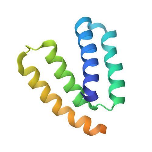
    The general stress response of Bacillus subtilis and close relatives provides the cell with protection from a variety of stresses. The upstream component of the environmental stress signal transduction cascade is activated by the RsbT kinase that switches binding partners from a 25 S macromolecular complex, the stressosome, to the RsbU phosphatase. Once the RsbU phosphatase is activated by interacting with RsbT, the alternative sigma factor, sigmaB, directs transcription of the general stress regulon. Previously, we demonstrated that the N-terminal domain of RsbU mediates the binding of RsbT. We now describe residues in N-RsbU that are crucial to this interaction by experimentation both in vitro and in vivo. Furthermore, crystal structures of the N-RsbU mutants provide a molecular explanation for the loss of interaction. Finally, we also characterize mutants in RsbT that affect binding to both RsbU and a simplified, binary model of the stressosome and thus identify overlapping binding surfaces on the RsbT "switch."


  • Organizational Affiliation

    Institute for Cell and Molecular Biosciences, Faculty of Medical Sciences, Newcastle University, Newcastle upon Tyne NE2 4HH, United Kingdom.


Macromolecules
Find similar proteins by:  (by identity cutoff)  |  3D Structure
Entity ID: 1
MoleculeChains Sequence LengthOrganismDetailsImage
PHOSPHOSERINE PHOSPHATASE RSBU
A, B, C, D, E
111Bacillus subtilisMutation(s): 1 
EC: 3.1.3.3
UniProt
Find proteins for P40399 (Bacillus subtilis (strain 168))
Explore P40399 
Go to UniProtKB:  P40399
Entity Groups  
Sequence Clusters30% Identity50% Identity70% Identity90% Identity95% Identity100% Identity
UniProt GroupP40399
Sequence Annotations
Expand
  • Reference Sequence
Experimental Data & Validation

Experimental Data

  • Method: X-RAY DIFFRACTION
  • Resolution: 1.85 Å
  • R-Value Free: 0.253 
  • R-Value Work: 0.183 
  • R-Value Observed: 0.186 
  • Space Group: C 1 2 1
Unit Cell:
Length ( Å )Angle ( ˚ )
a = 100.157α = 90
b = 47.831β = 104.36
c = 94.968γ = 90
Software Package:
Software NamePurpose
REFMACrefinement
MOSFLMdata reduction
SCALAdata scaling
ARP/wARPphasing

Structure Validation

View Full Validation Report


Entry History 

Deposition Data

Revision History  (Full details and data files)

  • Version 1.0: 2007-02-13
    Type: Initial release
  • Version 1.1: 2011-05-08
    Changes: Version format compliance
  • Version 1.2: 2011-07-13
    Changes: Version format compliance
  • Version 1.3: 2017-06-28
    Changes: Refinement description
  • Version 1.4: 2018-03-28
    Changes: Database references, Source and taxonomy
  • Version 1.5: 2023-12-13
    Changes: Advisory, Data collection, Database references, Refinement description