Structures of R- and T-State Escherichia Coli Aspartokinase III: Mechanisms of the Allosteric Transition and Inhibition by Lysine.
Kotaka, M., Ren, J., Lockyer, M., Hawkins, A.R., Stammers, D.K.(2006) J Biol Chem 281: 31544
- PubMed: 16905770
- DOI: https://doi.org/10.1074/jbc.M605886200
- Primary Citation of Related Structures:
2J0W, 2J0X - PubMed Abstract:
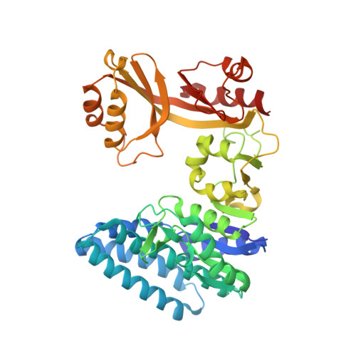
Aspartokinase III (AKIII) from Escherichia coli catalyzes an initial commitment step of the aspartate pathway, giving biosynthesis of certain amino acids including lysine. We report crystal structures of AKIII in the inactive T-state with bound feedback allosteric inhibitor lysine and in the R-state with aspartate and ADP. The structures reveal an unusual configuration for the regulatory ACT domains, in which ACT2 is inserted into ACT1 rather than the expected tandem repeat. Comparison of R- and T-state AKIII indicates that binding of lysine to the regulatory ACT1 domain in R-state AKIII instigates a series of changes that release a "latch", the beta15-alphaK loop, from the catalytic domain, which in turn undergoes large rotational rearrangements, promoting tetramer formation and completion of the transition to the T-state. Lysine-induced allosteric transition in AKIII involves both destabilizing the R-state and stabilizing the T-state tetramer. Rearrangement of the catalytic domain blocks the ATP-binding site, which is therefore the structural basis for allosteric inhibition of AKIII by lysine.
Organizational Affiliation:
Division of Structural Biology, The Wellcome Trust Centre for Human Genetics, University of Oxford, Roosevelt Drive, Oxford OX3 7BN, United Kingdom.