2IAE

Crystal structure of a protein phosphatase 2A (PP2A) holoenzyme.


Experimental Data Snapshot

  • Method: X-RAY DIFFRACTION
  • Resolution: 3.50 Å
  • R-Value Free: 0.316 
  • R-Value Work: 0.257 
  • R-Value Observed: 0.260 

wwPDB Validation   3D Report Full Report


This is version 2.0 of the entry. See complete history


Literature

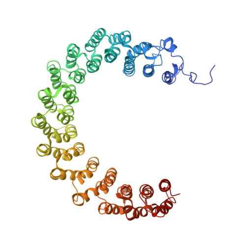
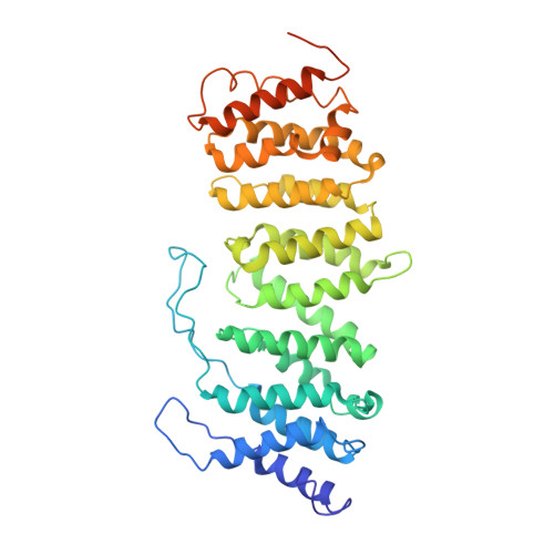
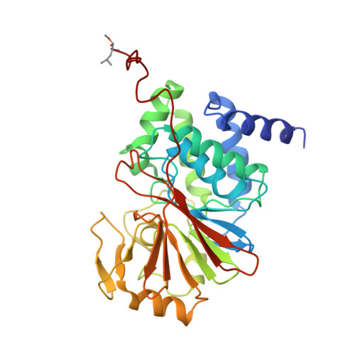
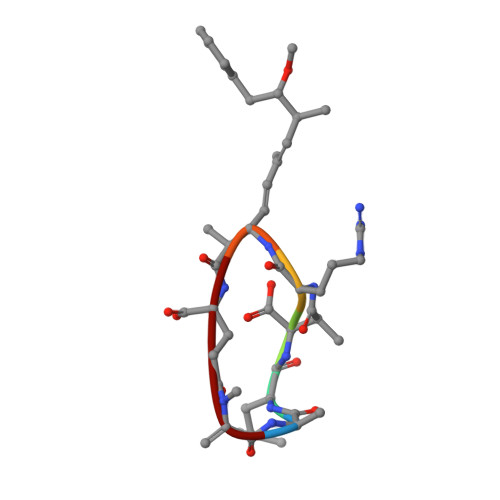
Crystal structure of a protein phosphatase 2A heterotrimeric holoenzyme.

Cho, U.S.Xu, W.

(2007) Nature 445: 53-57

  • DOI: https://doi.org/10.1038/nature05351
  • Primary Citation of Related Structures:  
    2IAE

  • PubMed Abstract: 

    Protein phosphatase 2A (PP2A) is a principal Ser/Thr phosphatase, the deregulation of which is associated with multiple human cancers, Alzheimer's disease and increased susceptibility to pathogen infections. How PP2A is structurally organized and functionally regulated remains unclear. Here we report the crystal structure of an AB'C heterotrimeric PP2A holoenzyme. The structure reveals that the HEAT repeats of the scaffold A subunit form a horseshoe-shaped fold, holding the catalytic C and regulatory B' subunits together on the same side. The regulatory B' subunit forms pseudo-HEAT repeats and interacts with the C subunit near the active site, thereby defining substrate specificity. The methylated carboxy-terminal tail of the C subunit interacts with a highly negatively charged region at the interface between A and B' subunits, suggesting that the C-terminal carboxyl methylation of the C subunit promotes B' subunit recruitment by neutralizing charge repulsion. Together, our structural results establish a crucial foundation for understanding PP2A assembly, substrate recruitment and regulation.


  • Organizational Affiliation

    Department of Biological Structure, University of Washington, Seattle, Washington 98195, USA.


Macromolecules
Find similar proteins by:  (by identity cutoff)  |  3D Structure
Entity ID: 1
MoleculeChains Sequence LengthOrganismDetailsImage
Serine/threonine-protein phosphatase 2A 65 kDa regulatory subunit A alpha isoform
A, D
589Mus musculusMutation(s): 0 
Gene Names: Ppp2r1a
EC: 3.1.3.16
UniProt
Find proteins for Q76MZ3 (Mus musculus)
Explore Q76MZ3 
Go to UniProtKB:  Q76MZ3
Entity Groups  
Sequence Clusters30% Identity50% Identity70% Identity90% Identity95% Identity100% Identity
UniProt GroupQ76MZ3
Sequence Annotations
Expand
  • Reference Sequence
Find similar proteins by:  (by identity cutoff)  |  3D Structure
Entity ID: 2
MoleculeChains Sequence LengthOrganismDetailsImage
Serine/threonine-protein phosphatase 2A 56 kDa regulatory subunit gamma isoform
B, E
407Homo sapiensMutation(s): 0 
Gene Names: PPP2R5CKIAA0044
EC: 3.1.3.16
UniProt & NIH Common Fund Data Resources
Find proteins for Q13362 (Homo sapiens)
Explore Q13362 
Go to UniProtKB:  Q13362
PHAROS:  Q13362
GTEx:  ENSG00000078304 
Entity Groups  
Sequence Clusters30% Identity50% Identity70% Identity90% Identity95% Identity100% Identity
UniProt GroupQ13362
Sequence Annotations
Expand
  • Reference Sequence
Find similar proteins by:  (by identity cutoff)  |  3D Structure
Entity ID: 3
MoleculeChains Sequence LengthOrganismDetailsImage
Serine/threonine-protein phosphatase 2A catalytic subunit alpha isoform
C, F
309Homo sapiensMutation(s): 1 
Gene Names: PPP2CA
EC: 3.1.3.16
UniProt & NIH Common Fund Data Resources
Find proteins for P67775 (Homo sapiens)
Explore P67775 
Go to UniProtKB:  P67775
PHAROS:  P67775
GTEx:  ENSG00000113575 
Entity Groups  
Sequence Clusters30% Identity50% Identity70% Identity90% Identity95% Identity100% Identity
UniProt GroupP67775
Sequence Annotations
Expand
  • Reference Sequence

Find similar proteins by:  Sequence   |   3D Structure  

Entity ID: 4
MoleculeChains Sequence LengthOrganismDetailsImage
microcystin-LRG [auth M],
H [auth N]
7Microcystis aeruginosaMutation(s): 0 
Sequence Annotations
Expand
  • Reference Sequence
Biologically Interesting Molecules (External Reference) 1 Unique
Experimental Data & Validation

Experimental Data

  • Method: X-RAY DIFFRACTION
  • Resolution: 3.50 Å
  • R-Value Free: 0.316 
  • R-Value Work: 0.257 
  • R-Value Observed: 0.260 
  • Space Group: P 21 3
Unit Cell:
Length ( Å )Angle ( ˚ )
a = 265.301α = 90
b = 265.301β = 90
c = 265.301γ = 90
Software Package:
Software NamePurpose
DENZOdata reduction
SCALEPACKdata scaling
SHARPphasing
DMphasing
REFMACrefinement
PDB_EXTRACTdata extraction
HKL-2000data collection
HKL-2000data reduction
HKL-2000data scaling

Structure Validation

View Full Validation Report


Entry History 

Deposition Data

Revision History  (Full details and data files)

  • Version 1.0: 2006-12-26
    Type: Initial release
  • Version 1.1: 2008-05-01
    Changes: Version format compliance
  • Version 1.2: 2011-07-13
    Changes: Advisory, Database references, Derived calculations, Non-polymer description, Version format compliance
  • Version 1.3: 2012-12-12
    Changes: Other
  • Version 1.4: 2017-10-18
    Changes: Advisory, Refinement description
  • Version 1.5: 2021-10-20
    Changes: Database references, Derived calculations
  • Version 2.0: 2023-11-15
    Changes: Atomic model, Data collection, Derived calculations