


2HYV
Human Annexin A2 with heparin hexasaccharide bound
- PDB DOI: https://doi.org/10.2210/pdb2HYV/pdb
- Classification: METAL BINDING PROTEIN
- Organism(s): Homo sapiens
- Expression System: Escherichia coli BL21(DE3)
- Mutation(s): No
- Deposited: 2006-08-07 Released: 2006-09-05
Experimental Data Snapshot
- Method: X-RAY DIFFRACTION
- Resolution: 1.42 Å
- R-Value Free: 0.223
- R-Value Work: 0.212
wwPDB Validation 3D Report Full Report

This is version 2.2 of the entry. See complete history.

Macromolecules
Find similar proteins by:
(by identity cutoff) | 3D Structure
Entity ID: 1 | |||||
|---|---|---|---|---|---|
| Molecule | Chains | Sequence Length | Organism | Details | Image |
| Annexin A2 | 308 | Homo sapiens | Mutation(s): 0 Gene Names: ANXA2 | 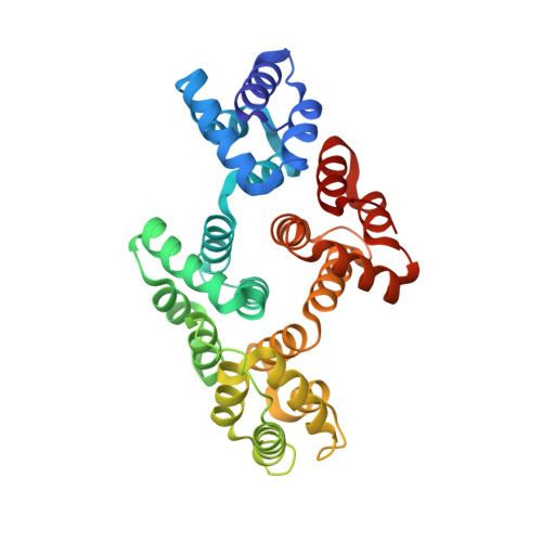 | |
UniProt & NIH Common Fund Data Resources | |||||
Find proteins for P07355 (Homo sapiens) Explore P07355 Go to UniProtKB: P07355 | |||||
PHAROS: P07355 GTEx: ENSG00000182718 | |||||
Entity Groups | |||||
| Sequence Clusters | 30% Identity50% Identity70% Identity90% Identity95% Identity100% Identity | ||||
| UniProt Group | P07355 | ||||
Sequence AnnotationsExpand | |||||
| |||||
Oligosaccharides
Entity ID: 2 | |||||
|---|---|---|---|---|---|
| Molecule | Chains | Length | 2D Diagram | Glycosylation | 3D Interactions |
| 4-deoxy-2-O-sulfo-alpha-L-threo-hex-4-enopyranuronic acid-(1-4)-2-deoxy-6-O-sulfo-2-(sulfoamino)-alpha-D-glucopyranose-(1-4)-2-O-sulfo-alpha-L-idopyranuronic acid-(1-4)-2-deoxy-6-O-sulfo-2-(sulfoamino)-alpha-D-glucopyranose-(1-4)-2-O-sulfo-alpha-L-idopyranuronic acid | B | 5 |  | N/A | |
Glycosylation Resources | |||||
GlyTouCan: G25610IB GlyCosmos: G25610IB | |||||
Small Molecules
| Ligands 1 Unique | |||||
|---|---|---|---|---|---|
| ID | Chains | Name / Formula / InChI Key | 2D Diagram | 3D Interactions | |
| CA Query on CA | C [auth A], D [auth A], E [auth A], F [auth A], G [auth A] | CALCIUM ION Ca BHPQYMZQTOCNFJ-UHFFFAOYSA-N |  | ||
Experimental Data & Validation
Experimental Data
- Method: X-RAY DIFFRACTION
- Resolution: 1.42 Å
- R-Value Free: 0.223
- R-Value Work: 0.212
- Space Group: P 21 21 2
Unit Cell:
| Length ( Å ) | Angle ( ˚ ) |
|---|---|
| a = 107.709 | α = 90 |
| b = 54.292 | β = 90 |
| c = 68.571 | γ = 90 |
| Software Name | Purpose |
|---|---|
| DENZO | data reduction |
| SCALEPACK | data scaling |
| EPMR | phasing |
| CNS | refinement |
| PDB_EXTRACT | data extraction |
Entry History
Deposition Data
- Released Date: 2006-09-05 Deposition Author(s): Shao, C., Head, J.F., Seaton, B.A.
Revision History (Full details and data files)
- Version 1.0: 2006-09-05
Type: Initial release - Version 1.1: 2008-05-01
Changes: Version format compliance - Version 1.2: 2011-07-13
Changes: Non-polymer description, Version format compliance - Version 1.3: 2017-10-18
Changes: Advisory, Refinement description - Version 2.0: 2020-07-29
Type: Remediation
Reason: Carbohydrate remediation
Changes: Advisory, Atomic model, Data collection, Derived calculations, Structure summary - Version 2.1: 2023-05-31
Changes: Database references, Derived calculations, Structure summary - Version 2.2: 2023-09-20
Changes: Data collection, Refinement description
