2HL6

Structure of homologously expressed Ferrulate esterase of Aspergillus niger in complex with CAPS


Experimental Data Snapshot

  • Method: X-RAY DIFFRACTION
  • Resolution: 1.55 Å
  • R-Value Free: 0.196 
  • R-Value Work: 0.163 
  • R-Value Observed: 0.165 

wwPDB Validation   3D Report Full Report


Ligand Structure Quality Assessment 


This is version 2.1 of the entry. See complete history


Literature

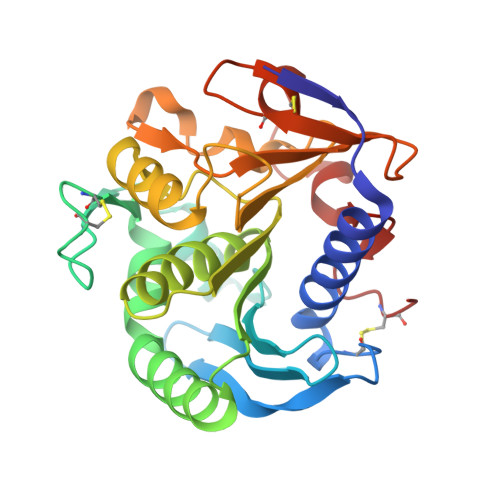
Respective importance of protein folding and glycosylation in the thermal stability of recombinant feruloyl esterase A

Benoit, I.Asther, M.Sulzenbacher, G.Record, E.Marmuse, L.Parsiegla, G.Herpoel-Gimbert, I.Asther, M.Bignon, C.

(2006) FEBS Lett 580: 5815-5821

  • DOI: https://doi.org/10.1016/j.febslet.2006.09.039
  • Primary Citation of Related Structures:  
    2HL6, 2IX9

  • PubMed Abstract: 

    The thermal stability of four molecular forms (native, refolded, glycosylated, non-glycosylated) of feruloyl esterase A (FAEA) was studied. From the most to the least thermo-resistant, the four molecular species ranked as follows: (i) glycosylated form produced native, (ii) non-glycosylated form produced native, (iii) non-glycosylated form produced as inclusion bodies and refolded, and (iv) glycosylated form produced native chemically denatured and then refolded. On the basis of these results and of crystal structure data, we discuss the respective importance of protein folding and glycosylation in the thermal stability of recombinant FAEA.


  • Organizational Affiliation

    UMR 1163 INRA/Universités de Provence et de la Méditerranée, Unité de Biotechnologie des Champignons Filamenteux, IFR86-BAIM, ESIL, 163 avenue de Luminy CP 925, 13288 MARSEILLE cedex 09, France.


Macromolecules
Find similar proteins by:  (by identity cutoff)  |  3D Structure
Entity ID: 1
MoleculeChains Sequence LengthOrganismDetailsImage
Feruloyl esterase A
A, B
260Aspergillus nigerMutation(s): 0 
Gene Names: faeA
EC: 3.1.1.73
UniProt
Find proteins for O42807 (Aspergillus niger)
Explore O42807 
Go to UniProtKB:  O42807
Entity Groups  
Sequence Clusters30% Identity50% Identity70% Identity90% Identity95% Identity100% Identity
UniProt GroupO42807
Sequence Annotations
Expand
  • Reference Sequence
Oligosaccharides

Help

Entity ID: 2
MoleculeChains Length2D Diagram Glycosylation3D Interactions
alpha-D-mannopyranose-(1-2)-alpha-D-mannopyranose-(1-3)-beta-D-mannopyranose-(1-4)-2-acetamido-2-deoxy-beta-D-glucopyranose-(1-4)-2-acetamido-2-deoxy-beta-D-glucopyranose
C
5N-Glycosylation
Glycosylation Resources
GlyTouCan:  G42227JK
GlyCosmos:  G42227JK
GlyGen:  G42227JK
Entity ID: 3
MoleculeChains Length2D Diagram Glycosylation3D Interactions
beta-D-mannopyranose-(1-4)-2-acetamido-2-deoxy-beta-D-glucopyranose-(1-4)-2-acetamido-2-deoxy-beta-D-glucopyranose
D
3N-Glycosylation
Glycosylation Resources
GlyTouCan:  G15407YE
GlyCosmos:  G15407YE
GlyGen:  G15407YE
Small Molecules
Ligands 3 Unique
IDChains Name / Formula / InChI Key2D Diagram3D Interactions
CXS
Query on CXS

Download Ideal Coordinates CCD File 
I [auth A],
R [auth B]
3-CYCLOHEXYL-1-PROPYLSULFONIC ACID
C9 H19 N O3 S
PJWWRFATQTVXHA-UHFFFAOYSA-N
SO4
Query on SO4

Download Ideal Coordinates CCD File 
E [auth A],
F [auth A],
J [auth B],
K [auth B]
SULFATE ION
O4 S
QAOWNCQODCNURD-UHFFFAOYSA-L
EDO
Query on EDO

Download Ideal Coordinates CCD File 
G [auth A]
H [auth A]
L [auth B]
M [auth B]
N [auth B]
G [auth A],
H [auth A],
L [auth B],
M [auth B],
N [auth B],
O [auth B],
P [auth B],
Q [auth B]
1,2-ETHANEDIOL
C2 H6 O2
LYCAIKOWRPUZTN-UHFFFAOYSA-N
Experimental Data & Validation

Experimental Data

  • Method: X-RAY DIFFRACTION
  • Resolution: 1.55 Å
  • R-Value Free: 0.196 
  • R-Value Work: 0.163 
  • R-Value Observed: 0.165 
  • Space Group: C 1 2 1
Unit Cell:
Length ( Å )Angle ( ˚ )
a = 145.52α = 90
b = 51.7β = 127.98
c = 99.588γ = 90
Software Package:
Software NamePurpose
SCALAdata scaling
AMoREphasing
REFMACrefinement
PDB_EXTRACTdata extraction
DENZOdata reduction
CCP4data scaling

Structure Validation

View Full Validation Report


Ligand Structure Quality Assessment 


Entry History 

Deposition Data

Revision History  (Full details and data files)

  • Version 1.0: 2006-10-31
    Type: Initial release
  • Version 1.1: 2008-05-01
    Changes: Version format compliance
  • Version 1.2: 2011-07-13
    Changes: Advisory, Version format compliance
  • Version 2.0: 2020-07-29
    Type: Remediation
    Reason: Carbohydrate remediation
    Changes: Atomic model, Data collection, Derived calculations, Structure summary
  • Version 2.1: 2023-10-25
    Changes: Data collection, Database references, Refinement description, Structure summary