2H8L

Crystal structure of the bb' fragment of ERp57


Experimental Data Snapshot

  • Method: X-RAY DIFFRACTION
  • Resolution: 2.00 Å
  • R-Value Free: 0.250 
  • R-Value Work: 0.194 
  • R-Value Observed: 0.197 

wwPDB Validation   3D Report Full Report


This is version 1.2 of the entry. See complete history


Literature

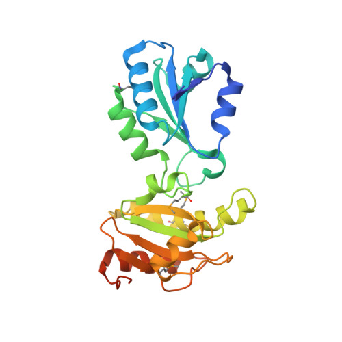
Crystal Structure of the bb' Domains of the Protein Disulfide Isomerase ERp57.

Kozlov, G.Maattanen, P.Schrag, J.D.Pollock, S.Cygler, M.Nagar, B.Thomas, D.Y.Gehring, K.

(2006) Structure 14: 1331-1339

  • DOI: https://doi.org/10.1016/j.str.2006.06.019
  • Primary Citation of Related Structures:  
    2H8L

  • PubMed Abstract: 

    The synthesis of proteins in the endoplasmic reticulum (ER) is limited by the rate of correct disulfide bond formation. This process is carried out by protein disulfide isomerases, a family of ER proteins which includes general enzymes such as PDI that recognize unfolded proteins and others that are selective for specific proteins or classes. Using small-angle X-ray scattering and X-ray crystallography, we report the structure of a selective isomerase, ERp57, and its interactions with the lectin chaperone calnexin. Using isothermal titration calorimetry and NMR spectroscopy, we show that the b' domain of ERp57 binds calnexin with micromolar affinity through a conserved patch of basic residues. Disruption of this binding site by mutagenesis abrogates folding of RNase B in an in vitro assay. The relative positions of the ERp57 catalytic sites and calnexin binding site suggest that activation by calnexin is due to substrate recruitment rather than a direct stimulation of ERp57 oxidoreductase activity.


  • Organizational Affiliation

    Biochemistry Department, McGill University, 3655 Promenade Sir William Osler, Montréal, Québec H3G 1Y6, Canada.


Macromolecules
Find similar proteins by:  (by identity cutoff)  |  3D Structure
Entity ID: 1
MoleculeChains Sequence LengthOrganismDetailsImage
Protein disulfide-isomerase A3
A, B, C
252Homo sapiensMutation(s): 5 
Gene Names: PDIA3ERP60GRP58
EC: 5.3.4.1
UniProt & NIH Common Fund Data Resources
Find proteins for P30101 (Homo sapiens)
Explore P30101 
Go to UniProtKB:  P30101
PHAROS:  P30101
GTEx:  ENSG00000167004 
Entity Groups  
Sequence Clusters30% Identity50% Identity70% Identity90% Identity95% Identity100% Identity
UniProt GroupP30101
Sequence Annotations
Expand
  • Reference Sequence
Small Molecules
Modified Residues  1 Unique
IDChains TypeFormula2D DiagramParent
MSE
Query on MSE
A, B, C
L-PEPTIDE LINKINGC5 H11 N O2 SeMET
Experimental Data & Validation

Experimental Data

  • Method: X-RAY DIFFRACTION
  • Resolution: 2.00 Å
  • R-Value Free: 0.250 
  • R-Value Work: 0.194 
  • R-Value Observed: 0.197 
  • Space Group: P 1 21 1
Unit Cell:
Length ( Å )Angle ( ˚ )
a = 76.34α = 90
b = 62.415β = 98.4
c = 99.293γ = 90
Software Package:
Software NamePurpose
REFMACrefinement
ADSCdata collection
HKL-2000data scaling
SHELXSphasing

Structure Validation

View Full Validation Report


Entry History 

Deposition Data

Revision History  (Full details and data files)

  • Version 1.0: 2006-08-29
    Type: Initial release
  • Version 1.1: 2008-05-01
    Changes: Version format compliance
  • Version 1.2: 2011-07-13
    Changes: Advisory, Version format compliance