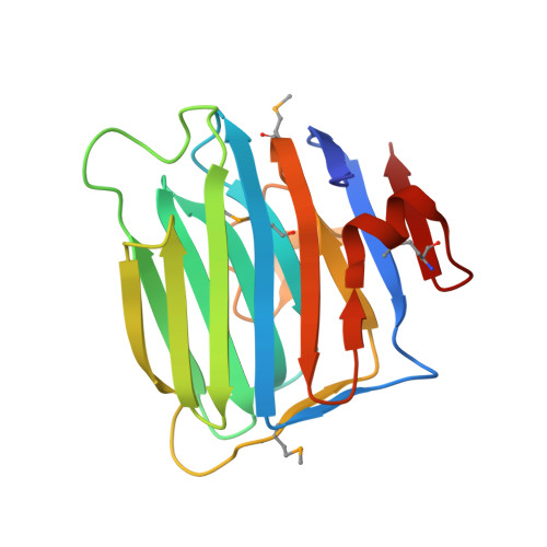
Crystal Structure of the Second LNS/LG Domain from Neurexin 1{alpha}: Ca2+ binding and the effects of alternative splicing
Sheckler, L.R., Henry, L., Sugita, S., Sudhof, T.C., Rudenko, G.(2006) J Biol Chem 281: 22896-22905
- PubMed: 16772286
- DOI: https://doi.org/10.1074/jbc.M603464200
- Primary Citation of Related Structures:
2H0B - PubMed Abstract:
Neurexins mediate protein interactions at the synapse, playing an essential role in synaptic function. Extracellular domains of neurexins, and their fragments, bind a distinct profile of different proteins regulated by alternative splicing and Ca2+. The crystal structure of n1alpha_LNS#2 (the second LNS/LG domain of bovine neurexin 1alpha) reveals large structural differences compared with n1alpha_LNS#6 (or n1beta_LNS), the only other LNS/LG domain for which a structure has been determined. The differences overlap the so-called hyper-variable surface, the putative protein interaction surface that is reshaped as a result of alternative splicing. A Ca2+-binding site is revealed at the center of the hyper-variable surface next to splice insertion sites. Isothermal titration calorimetry indicates that the Ca2+-binding site in n1alpha_LNS#2 has low affinity (Kd approximately 400 microm). Ca2+ binding ceases to be measurable when an 8- or 15-residue splice insert is present at the splice site SS#2 indicating that alternative splicing can affect Ca2+-binding sites of neurexin LNS/LG domains. Our studies initiate a framework for the putative protein interaction sites of neurexin LNS/LG domains. This framework is essential to understand how incorporation of alternative splice inserts expands the information from a limited set of neurexin genes to produce a large array of synaptic adhesion molecules with potentially very different synaptic function.
Organizational Affiliation:
Life Sciences Institute and Department of Pharmacology, The University of Michigan, Ann Arbor, Michigan 48109-2216, USA.