2GUX

Selenomethionine derivative of griffithsin


Experimental Data Snapshot

  • Method: X-RAY DIFFRACTION
  • Resolution: 2.00 Å
  • R-Value Free: 0.254 
  • R-Value Work: 0.189 
  • R-Value Observed: 0.189 

wwPDB Validation   3D Report Full Report


This is version 1.2 of the entry. See complete history


Literature

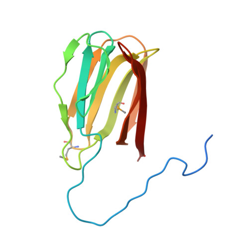
Domain-swapped structure of the potent antiviral protein griffithsin and its mode of carbohydrate binding.

Ziolkowska, N.E.O'keefe, B.R.Mori, T.Zhu, C.Giomarelli, B.Vojdani, F.Palmer, K.E.McMahon, J.B.Wlodawer, A.

(2006) Structure 14: 1127-1135

  • DOI: https://doi.org/10.1016/j.str.2006.05.017
  • Primary Citation of Related Structures:  
    2GTY, 2GUC, 2GUD, 2GUE, 2GUX

  • PubMed Abstract: 

    The crystal structure of griffithsin, an antiviral lectin from the red alga Griffithsia sp., was solved and refined at 1.3 A resolution for the free protein and 0.94 A for a complex with mannose. Griffithsin molecules form a domain-swapped dimer, in which two beta strands of one molecule complete a beta prism consisting of three four-stranded sheets, with an approximate 3-fold axis, of another molecule. The structure of each monomer bears close resemblance to jacalin-related lectins, but its dimeric structure is unique. The structures of complexes of griffithsin with mannose and N-acetylglucosamine defined the locations of three almost identical carbohydrate binding sites on each monomer. We have also shown that griffithsin is a potent inhibitor of the coronavirus responsible for severe acute respiratory syndrome (SARS). Antiviral potency of griffithsin is likely due to the presence of multiple, similar sugar binding sites that provide redundant attachment points for complex carbohydrate molecules present on viral envelopes.


  • Organizational Affiliation

    Protein Structure Section, Macromolecular Crystallography Laboratory, National Cancer Institute, NCI-Frederick, Frederick, MD 21702, USA.


Macromolecules
Find similar proteins by:  (by identity cutoff)  |  3D Structure
Entity ID: 1
MoleculeChains Sequence LengthOrganismDetailsImage
griffithsin138GriffithsiaMutation(s): 2 
UniProt
Find proteins for P84801 (Griffithsia sp. (strain Q66D336))
Explore P84801 
Go to UniProtKB:  P84801
Entity Groups  
Sequence Clusters30% Identity50% Identity70% Identity90% Identity95% Identity100% Identity
UniProt GroupP84801
Sequence Annotations
Expand
  • Reference Sequence
Small Molecules
Ligands 1 Unique
IDChains Name / Formula / InChI Key2D Diagram3D Interactions
SO4
Query on SO4

Download Ideal Coordinates CCD File 
B [auth A]SULFATE ION
O4 S
QAOWNCQODCNURD-UHFFFAOYSA-L
Modified Residues  1 Unique
IDChains TypeFormula2D DiagramParent
MSE
Query on MSE
A
L-PEPTIDE LINKINGC5 H11 N O2 SeMET
Experimental Data & Validation

Experimental Data

  • Method: X-RAY DIFFRACTION
  • Resolution: 2.00 Å
  • R-Value Free: 0.254 
  • R-Value Work: 0.189 
  • R-Value Observed: 0.189 
  • Space Group: P 32 2 1
Unit Cell:
Length ( Å )Angle ( ˚ )
a = 66.87α = 90
b = 66.87β = 90
c = 68.2γ = 120
Software Package:
Software NamePurpose
REFMACrefinement
HKL-2000data reduction
DENZOdata reduction
HKL-2000data scaling
SCALEPACKdata scaling
HKL-3000phasing
SHELXEmodel building
SOLVEphasing
RESOLVEphasing
ARP/wARPmodel building

Structure Validation

View Full Validation Report


Entry History 

Deposition Data

Revision History  (Full details and data files)

  • Version 1.0: 2006-08-01
    Type: Initial release
  • Version 1.1: 2008-05-01
    Changes: Version format compliance
  • Version 1.2: 2011-07-13
    Changes: Advisory, Derived calculations, Version format compliance