2FU3

Crystal structure of gephyrin E-domain


Experimental Data Snapshot

  • Method: X-RAY DIFFRACTION
  • Resolution: 2.70 Å
  • R-Value Free: 0.273 
  • R-Value Work: 0.202 

wwPDB Validation   3D Report Full Report


This is version 1.4 of the entry. See complete history


Literature

Deciphering the structural framework of glycine receptor anchoring by gephyrin.

Kim, E.Y.Schrader, N.Smolinsky, B.Bedet, C.Vannier, C.Schwarz, G.Schindelin, H.

(2006) EMBO J 25: 1385-1395

  • DOI: https://doi.org/10.1038/sj.emboj.7601029
  • Primary Citation of Related Structures:  
    2FTS, 2FU3

  • PubMed Abstract: 

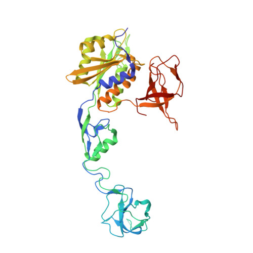
    Glycine is the major inhibitory neurotransmitter in the spinal cord and brain stem. Gephyrin is required to achieve a high concentration of glycine receptors (GlyRs) in the postsynaptic membrane, which is crucial for efficient glycinergic signal transduction. The interaction between gephyrin and the GlyR involves the E-domain of gephyrin and a cytoplasmic loop located between transmembrane segments three and four of the GlyR beta subunit. Here, we present crystal structures of the gephyrin E-domain with and without the GlyR beta-loop at 2.4 and 2.7 A resolutions, respectively. The GlyR beta-loop is bound in a symmetric 'key and lock' fashion to each E-domain monomer in a pocket adjacent to the dimer interface. Structure-guided mutagenesis followed by in vitro binding and in vivo colocalization assays demonstrate that a hydrophobic interaction formed by Phe 330 of gephyrin and Phe 398 and Ile 400 of the GlyR beta-loop is crucial for binding.


  • Organizational Affiliation

    Department of Biochemistry, Center for Structural Biology, State University of New York, Stony Brook, NY 11794-5115, USA.


Macromolecules
Find similar proteins by:  (by identity cutoff)  |  3D Structure
Entity ID: 1
MoleculeChains Sequence LengthOrganismDetailsImage
gephyrin
A, B
419Rattus norvegicusMutation(s): 0 
Gene Names: Rattus norvegicus (Norway rat)
UniProt
Find proteins for Q03555 (Rattus norvegicus)
Explore Q03555 
Go to UniProtKB:  Q03555
Entity Groups  
Sequence Clusters30% Identity50% Identity70% Identity90% Identity95% Identity100% Identity
UniProt GroupQ03555
Sequence Annotations
Expand
  • Reference Sequence
Experimental Data & Validation

Experimental Data

  • Method: X-RAY DIFFRACTION
  • Resolution: 2.70 Å
  • R-Value Free: 0.273 
  • R-Value Work: 0.202 
  • Space Group: P 21 21 2
Unit Cell:
Length ( Å )Angle ( ˚ )
a = 108.82α = 90
b = 156.195β = 90
c = 51.362γ = 90
Software Package:
Software NamePurpose
DENZOdata reduction
SCALEPACKdata scaling
MOLREPphasing
REFMACrefinement
PDB_EXTRACTdata extraction
HKL-2000data reduction

Structure Validation

View Full Validation Report


Entry History 

Deposition Data

Revision History  (Full details and data files)

  • Version 1.0: 2006-03-14
    Type: Initial release
  • Version 1.1: 2008-05-01
    Changes: Version format compliance
  • Version 1.2: 2011-07-13
    Changes: Advisory, Version format compliance
  • Version 1.3: 2017-10-18
    Changes: Refinement description
  • Version 1.4: 2024-02-14
    Changes: Data collection, Database references