2FJU

Activated Rac1 bound to its effector phospholipase C beta 2


Experimental Data Snapshot

  • Method: X-RAY DIFFRACTION
  • Resolution: 2.20 Å
  • R-Value Free: 0.226 
  • R-Value Work: 0.208 

wwPDB Validation   3D Report Full Report


Ligand Structure Quality Assessment 


This is version 1.3 of the entry. See complete history


Literature

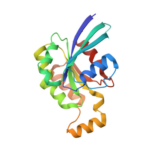
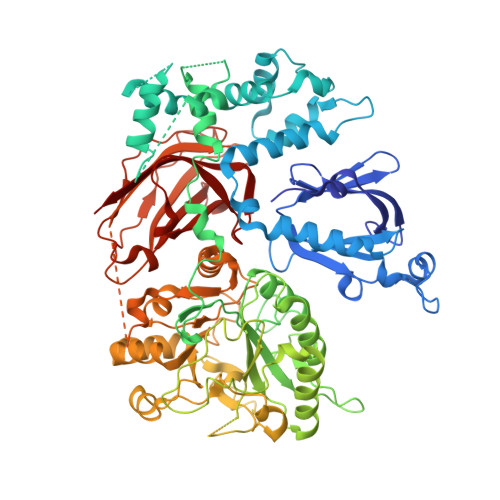
Crystal structure of Rac1 bound to its effector phospholipase C-beta2.

Jezyk, M.R.Snyder, J.T.Gershberg, S.Worthylake, D.K.Harden, T.K.Sondek, J.

(2006) Nat Struct Mol Biol 13: 1135-1140

  • DOI: https://doi.org/10.1038/nsmb1175
  • Primary Citation of Related Structures:  
    2FJU

  • PubMed Abstract: 

    Although diverse signaling cascades require the coordinated regulation of heterotrimeric G proteins and small GTPases, these connections remain poorly understood. We present the crystal structure of the GTPase Rac1 bound to phospholipase C-beta2 (PLC-beta2), a classic effector of heterotrimeric G proteins. Rac1 engages the pleckstrin-homology (PH) domain of PLC-beta2 to optimize its orientation for substrate membranes. Gbetagamma also engages the PH domain to activate PLC-beta2, and these two activation events are compatible, leading to additive stimulation of phospholipase activity. In contrast to PLC-delta, the PH domain of PLC-beta2 cannot bind phosphoinositides, eliminating this mode of regulation. The structure of the Rac1-PLC-beta2 complex reveals determinants that dictate selectivity of PLC-beta isozymes for Rac GTPases over other Rho-family GTPases, and substitutions within PLC-beta2 abrogate its stimulation by Rac1 but not by Gbetagamma, allowing for functional dissection of this integral signaling node.


  • Organizational Affiliation

    Department of Biochemistry and Biophysics The University of North Carolina School of Medicine, Chapel Hill, North Carolina 27599, USA.


Macromolecules
Find similar proteins by:  (by identity cutoff)  |  3D Structure
Entity ID: 1
MoleculeChains Sequence LengthOrganismDetailsImage
Ras-related C3 botulinum toxin substrate 1178Homo sapiensMutation(s): 0 
Gene Names: RAC1
UniProt & NIH Common Fund Data Resources
Find proteins for P63000 (Homo sapiens)
Explore P63000 
Go to UniProtKB:  P63000
PHAROS:  P63000
GTEx:  ENSG00000136238 
Entity Groups  
Sequence Clusters30% Identity50% Identity70% Identity90% Identity95% Identity100% Identity
UniProt GroupP63000
Sequence Annotations
Expand
  • Reference Sequence
Find similar proteins by:  (by identity cutoff)  |  3D Structure
Entity ID: 2
MoleculeChains Sequence LengthOrganismDetailsImage
1-phosphatidylinositol-4,5-bisphosphate phosphodiesterase beta 2799Homo sapiensMutation(s): 0 
Gene Names: PLCB2
EC: 3.1.4.11
UniProt & NIH Common Fund Data Resources
Find proteins for Q00722 (Homo sapiens)
Explore Q00722 
Go to UniProtKB:  Q00722
PHAROS:  Q00722
GTEx:  ENSG00000137841 
Entity Groups  
Sequence Clusters30% Identity50% Identity70% Identity90% Identity95% Identity100% Identity
UniProt GroupQ00722
Sequence Annotations
Expand
  • Reference Sequence
Experimental Data & Validation

Experimental Data

  • Method: X-RAY DIFFRACTION
  • Resolution: 2.20 Å
  • R-Value Free: 0.226 
  • R-Value Work: 0.208 
  • Space Group: P 32 2 1
Unit Cell:
Length ( Å )Angle ( ˚ )
a = 185.822α = 90
b = 185.822β = 90
c = 93.823γ = 120
Software Package:
Software NamePurpose
HKL-2000data collection
SCALEPACKdata scaling
SHELXmodel building
MLPHAREphasing
DMmodel building
CNSrefinement
HKL-2000data reduction
SHELXphasing
DMphasing

Structure Validation

View Full Validation Report


Ligand Structure Quality Assessment 


Entry History 

Deposition Data

Revision History  (Full details and data files)

  • Version 1.0: 2006-11-21
    Type: Initial release
  • Version 1.1: 2008-05-01
    Changes: Version format compliance
  • Version 1.2: 2011-07-13
    Changes: Version format compliance
  • Version 1.3: 2024-02-14
    Changes: Data collection, Database references, Derived calculations