Structural Characterization of the Split Pleckstrin Homology Domain in Phospholipase C-{gamma}1 and Its Interaction with TRPC3
Wen, W., Yan, J., Zhang, M.(2006) J Biol Chem 281: 12060-12068
- PubMed: 16500902
- DOI: https://doi.org/10.1074/jbc.M600336200
- Primary Citation of Related Structures:
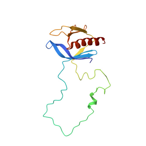
2FJL - PubMed Abstract:
Phospholipase C (PLC)-gamma is unique among the PLC enzymes because each PLC-gamma isozyme contains a split pleckstrin homology (PH) domain with an SH2SH2SH3 tandem repeat insertion (where SH indicates Src homology domain) in the middle of its sequence. Split PH domains exist in a number of other proteins that play crucial signaling roles. However, little is known about the structure and function of split PH domains. The C-terminal half of the PLC-gamma split PH domain has been implicated to interact directly with the TRPC3 calcium channel, thereby providing a direct coupling mechanism between PLC-gamma and agonist-induced calcium entry. However, this interaction has not been proved by direct biochemical or structural studies. Here we determined the three-dimensional structure of the split PH domain of PLC-gamma1, and we found that the split PH domain of the enzyme folds into a canonical PH domain fold with high thermostability. The SH2SH2SH3 insertion between the beta3 and beta4 strands does not change the structure of the split PH domain. In contrast to the majority of phospholipid-binding PH domains, the PLC-gamma1 split PH domain lacks the signature lipid-binding motif located between the beta1 and beta2 strands. Consistent with this structural feature, the split PH domain of PLC-gamma1 does not bind to phospholipids. Multiple biochemical and biophysical experiments have argued against a direct interaction between TRPC3 and the C-terminal half of the PLC-gamma1 split PH domain. Our data pointed to the existence of a yet to be elucidated interaction mechanism between TRPC3 and PLC-gamma1.
Organizational Affiliation:
Department of Biochemistry, Molecular Neuroscience Center, Hong Kong University of Science and Technology, Clear Water Bay, Kowloon, Hong Kong, China.