2FFF

Open Form of a Class A Transpeptidase Domain


Experimental Data Snapshot

  • Method: X-RAY DIFFRACTION
  • Resolution: 2.23 Å
  • R-Value Free: 0.246 
  • R-Value Work: 0.183 
  • R-Value Observed: 0.186 

wwPDB Validation   3D Report Full Report


This is version 1.3 of the entry. See complete history


Literature

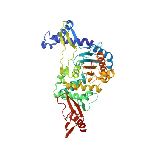
Structural analysis of an "open" form of PBP1B from Streptococcus pneumoniae.

Lovering, A.L.De Castro, L.Lim, D.Strynadka, N.C.

(2006) Protein Sci 15: 1701-1709

  • DOI: https://doi.org/10.1110/ps.062112106
  • Primary Citation of Related Structures:  
    2FFF

  • PubMed Abstract: 

    The class A PBP1b from Streptococcus pneumoniae is responsible for glycosyltransferase and transpeptidase (TP) reactions, forming the peptidoglycan of the bacterial cell wall. The enzyme has been produced in a stable, soluble form and undergoes time-dependent proteolysis to leave an intact TP domain. Crystals of this TP domain were obtained, diffracting to 2.2 A resolution, and the structure was solved by using molecular replacement. Analysis of the structure revealed an "open" active site, with important conformational differences to the previously determined "closed" apoenzyme. The active-site nucleophile, Ser460, is in an orientation that allows for acylation by beta-lactams. Consistent with the productive conformation of the conserved active-site catalytic residues, adjacent loops show only minor deviation from those of known acyl-enzyme structures. These findings are discussed in the context of enzyme functionality and the possible conformational sampling of PBP1b between active and inactive states.


  • Organizational Affiliation

    Department of Biochemistry and Molecular Biology, Center for Blood Research, University of British Columbia, Vancouver, Canada.


Macromolecules

Find similar proteins by:  Sequence   |   3D Structure  

Entity ID: 1
MoleculeChains Sequence LengthOrganismDetailsImage
penicillin-binding protein 1B15Streptococcus pneumoniaeMutation(s): 0 
UniProt
Find proteins for Q4TUQ1 (Streptococcus pneumoniae)
Explore Q4TUQ1 
Go to UniProtKB:  Q4TUQ1
Entity Groups  
Sequence Clusters30% Identity50% Identity70% Identity90% Identity95% Identity100% Identity
UniProt GroupQ4TUQ1
Sequence Annotations
Expand
  • Reference Sequence
Find similar proteins by:  (by identity cutoff)  |  3D Structure
Entity ID: 2
MoleculeChains Sequence LengthOrganismDetailsImage
penicillin-binding protein 1B453Streptococcus pneumoniaeMutation(s): 0 
UniProt
Find proteins for O70038 (Streptococcus pneumoniae)
Explore O70038 
Go to UniProtKB:  O70038
Entity Groups  
Sequence Clusters30% Identity50% Identity70% Identity90% Identity95% Identity100% Identity
UniProt GroupO70038
Sequence Annotations
Expand
  • Reference Sequence
Small Molecules
Ligands 1 Unique
IDChains Name / Formula / InChI Key2D Diagram3D Interactions
NI
Query on NI

Download Ideal Coordinates CCD File 
C [auth B]NICKEL (II) ION
Ni
VEQPNABPJHWNSG-UHFFFAOYSA-N
Experimental Data & Validation

Experimental Data

  • Method: X-RAY DIFFRACTION
  • Resolution: 2.23 Å
  • R-Value Free: 0.246 
  • R-Value Work: 0.183 
  • R-Value Observed: 0.186 
  • Space Group: C 1 2 1
Unit Cell:
Length ( Å )Angle ( ˚ )
a = 131.661α = 90
b = 78.043β = 113.02
c = 60.315γ = 90
Software Package:
Software NamePurpose
REFMACrefinement
PDB_EXTRACTdata extraction
MOSFLMdata reduction
CCP4data scaling
MOLREPphasing

Structure Validation

View Full Validation Report


Entry History 

Deposition Data

Revision History  (Full details and data files)

  • Version 1.0: 2006-06-20
    Type: Initial release
  • Version 1.1: 2008-05-01
    Changes: Version format compliance
  • Version 1.2: 2011-07-13
    Changes: Advisory, Version format compliance
  • Version 1.3: 2023-08-30
    Changes: Data collection, Database references, Derived calculations, Refinement description