2F6H

Myosin V cargo binding domain


Experimental Data Snapshot

  • Method: X-RAY DIFFRACTION
  • Resolution: 2.25 Å
  • R-Value Free: 0.250 
  • R-Value Work: 0.217 
  • R-Value Observed: 0.219 

wwPDB Validation   3D Report Full Report


This is version 1.3 of the entry. See complete history


Literature

Structural basis for myosin V discrimination between distinct cargoes

Pashkova, N.Jin, Y.Ramaswamy, S.Weisman, L.S.

(2006) EMBO J 25: 693-700

  • DOI: https://doi.org/10.1038/sj.emboj.7600965
  • Primary Citation of Related Structures:  
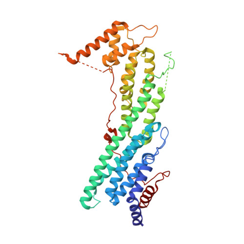
    2F6H

  • PubMed Abstract: 

    Myosin V molecular motors move cargoes on actin filaments. A myosin V may move multiple cargoes to distinct places at different times. The cargoes attach to the globular tail of myosin V via cargo-specific receptors. Here we report the crystal structure at 2.2 A of the myosin V globular tail. The overall tertiary structure has not been previously observed. There are several patches of highly conserved regions distributed on the surface of the tail. These are candidate attachment sites for cargo-specific receptors. Indeed, we identified a region of five conserved surface residues that are solely required for vacuole inheritance. Likewise, we identified a region of five conserved surface residues that are required for secretory vesicle movement, but not vacuole movement. These two regions are at opposite ends of the oblong-shaped cargo-binding domain, and moreover are offset by 180 degrees. The fact that the cargo-binding areas are distant from each other and simultaneously exposed on the surface of the globular tail suggests that major targets for the regulation of cargo attachment are organelle-specific myosin V receptors.


  • Organizational Affiliation

    Department of Biochemistry, University of Iowa, Iowa City, IA 52242, USA.


Macromolecules
Find similar proteins by:  (by identity cutoff)  |  3D Structure
Entity ID: 1
MoleculeChains Sequence LengthOrganismDetailsImage
Myosin-2A [auth X]419Saccharomyces cerevisiaeMutation(s): 0 
Gene Names: MYO2CDC66
UniProt
Find proteins for P19524 (Saccharomyces cerevisiae (strain ATCC 204508 / S288c))
Explore P19524 
Go to UniProtKB:  P19524
Entity Groups  
Sequence Clusters30% Identity50% Identity70% Identity90% Identity95% Identity100% Identity
UniProt GroupP19524
Sequence Annotations
Expand
  • Reference Sequence
Experimental Data & Validation

Experimental Data

  • Method: X-RAY DIFFRACTION
  • Resolution: 2.25 Å
  • R-Value Free: 0.250 
  • R-Value Work: 0.217 
  • R-Value Observed: 0.219 
  • Space Group: P 21 21 21
Unit Cell:
Length ( Å )Angle ( ˚ )
a = 50.78α = 90
b = 72.673β = 90
c = 167.982γ = 90
Software Package:
Software NamePurpose
d*TREKdata reduction
SOLVEphasing
REFMACrefinement
d*TREKdata scaling

Structure Validation

View Full Validation Report


Entry History 

Deposition Data

Revision History  (Full details and data files)

  • Version 1.0: 2006-02-07
    Type: Initial release
  • Version 1.1: 2008-05-01
    Changes: Version format compliance
  • Version 1.2: 2011-07-13
    Changes: Version format compliance
  • Version 1.3: 2024-02-14
    Changes: Data collection, Database references