2EY4

Crystal Structure of a Cbf5-Nop10-Gar1 Complex


Experimental Data Snapshot

  • Method: X-RAY DIFFRACTION
  • Resolution: 2.11 Å
  • R-Value Free: 0.255 
  • R-Value Work: 0.211 
  • R-Value Observed: 0.211 

wwPDB Validation   3D Report Full Report


This is version 1.4 of the entry. See complete history


Literature

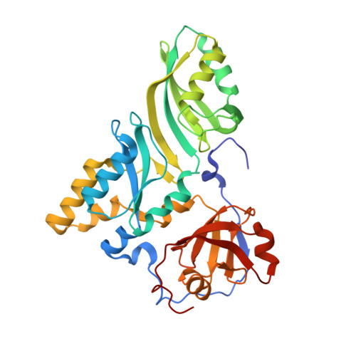
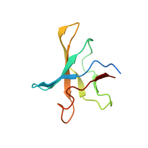
Crystal structure of a Cbf5-Nop10-Gar1 complex and implications in RNA-guided pseudouridylation and dyskeratosis congenita.

Rashid, R.Liang, B.Baker, D.L.Youssef, O.A.He, Y.Phipps, K.Terns, R.M.Terns, M.P.Li, H.

(2006) Mol Cell 21: 249-260

  • DOI: https://doi.org/10.1016/j.molcel.2005.11.017
  • Primary Citation of Related Structures:  
    2EY4

  • PubMed Abstract: 

    H/ACA RNA-protein complexes, comprised of four proteins and an H/ACA guide RNA, modify ribosomal and small nuclear RNAs. The H/ACA proteins are also essential components of telomerase in mammals. Cbf5 is the H/ACA protein that catalyzes isomerization of uridine to pseudouridine in target RNAs. Mutations in human Cbf5 (dyskerin) lead to dyskeratosis congenita. Here, we describe the 2.1 A crystal structure of a specific complex of three archaeal H/ACA proteins, Cbf5, Nop10, and Gar1. Cbf5 displays structural properties that are unique among known pseudouridine synthases and are consistent with its distinct function in RNA-guided pseudouridylation. We also describe the previously unknown structures of both Nop10 and Gar1 and the structural basis for their essential roles in pseudouridylation. By using information from related structures, we have modeled the entire ribonucleoprotein complex including both guide and substrate RNAs. We have also identified a dyskeratosis congenita mutation cluster site within a modeled dyskerin structure.


  • Organizational Affiliation

    Department of Chemistry and Biochemistry, Institute of Molecular Biophysics, Florida State University, Tallahassee, Florida 32306, USA.


Macromolecules
Find similar proteins by:  (by identity cutoff)  |  3D Structure
Entity ID: 1
MoleculeChains Sequence LengthOrganismDetailsImage
Probable tRNA pseudouridine synthase B
A, B
333Pyrococcus furiosusMutation(s): 0 
Gene Names: truB
EC: 5.4.99
UniProt
Find proteins for Q7LWY0 (Pyrococcus furiosus (strain ATCC 43587 / DSM 3638 / JCM 8422 / Vc1))
Explore Q7LWY0 
Go to UniProtKB:  Q7LWY0
Entity Groups  
Sequence Clusters30% Identity50% Identity70% Identity90% Identity95% Identity100% Identity
UniProt GroupQ7LWY0
Sequence Annotations
Expand
  • Reference Sequence
Find similar proteins by:  (by identity cutoff)  |  3D Structure
Entity ID: 2
MoleculeChains Sequence LengthOrganismDetailsImage
small nucleolar rnp similar to gar1
C, D
82Pyrococcus furiosus DSM 3638Mutation(s): 0 
UniProt
Find proteins for Q8U029 (Pyrococcus furiosus (strain ATCC 43587 / DSM 3638 / JCM 8422 / Vc1))
Explore Q8U029 
Go to UniProtKB:  Q8U029
Entity Groups  
Sequence Clusters30% Identity50% Identity70% Identity90% Identity95% Identity100% Identity
UniProt GroupQ8U029
Sequence Annotations
Expand
  • Reference Sequence
Find similar proteins by:  (by identity cutoff)  |  3D Structure
Entity ID: 3
MoleculeChains Sequence LengthOrganismDetailsImage
Ribosome biogenesis protein Nop10
E, F
55Pyrococcus furiosusMutation(s): 0 
UniProt
Find proteins for Q8U1R4 (Pyrococcus furiosus (strain ATCC 43587 / DSM 3638 / JCM 8422 / Vc1))
Explore Q8U1R4 
Go to UniProtKB:  Q8U1R4
Entity Groups  
Sequence Clusters30% Identity50% Identity70% Identity90% Identity95% Identity100% Identity
UniProt GroupQ8U1R4
Sequence Annotations
Expand
  • Reference Sequence
Experimental Data & Validation

Experimental Data

  • Method: X-RAY DIFFRACTION
  • Resolution: 2.11 Å
  • R-Value Free: 0.255 
  • R-Value Work: 0.211 
  • R-Value Observed: 0.211 
  • Space Group: P 1
Unit Cell:
Length ( Å )Angle ( ˚ )
a = 36.087α = 95.9
b = 75.361β = 96.9
c = 103.24γ = 94.99
Software Package:
Software NamePurpose
CNSrefinement
bioteXdata reduction
CNSphasing

Structure Validation

View Full Validation Report


Entry History 

Deposition Data

Revision History  (Full details and data files)

  • Version 1.0: 2006-01-24
    Type: Initial release
  • Version 1.1: 2008-05-01
    Changes: Version format compliance
  • Version 1.2: 2011-07-13
    Changes: Source and taxonomy, Version format compliance
  • Version 1.3: 2017-10-18
    Changes: Refinement description
  • Version 1.4: 2024-02-14
    Changes: Data collection, Database references, Derived calculations