2ESF

Identification of a Novel Non-Catalytic Bicarbonate Binding Site in Eubacterial beta-Carbonic Anhydrase


Experimental Data Snapshot

  • Method: X-RAY DIFFRACTION
  • Resolution: 2.25 Å
  • R-Value Free: 0.228 
  • R-Value Work: 0.198 
  • R-Value Observed: 0.199 

wwPDB Validation   3D Report Full Report


This is version 1.3 of the entry. See complete history


Literature

Identification of a Novel Noncatalytic Bicarbonate Binding Site in Eubacterial beta-Carbonic Anhydrase.

Cronk, J.D.Rowlett, R.S.Zhang, K.Y.J.Tu, C.Endrizzi, J.A.Lee, J.Gareiss, P.C.Preiss, J.R.

(2006) Biochemistry 45: 4351-4361

  • DOI: https://doi.org/10.1021/bi052272q
  • Primary Citation of Related Structures:  
    2A8C, 2A8D, 2ESF

  • PubMed Abstract: 

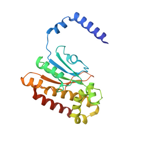
    The structures of beta class carbonic anhydrases (beta-CAs) determined so far fall into two distinct subclasses based on the observed coordination of the catalytic zinc (Zn2+) ion. The subclass of beta-CAs that coordinate Zn2+ tetrahedrally with four protein-derived ligands is represented by the structures of orthologues from Porphyridium purpureum, Escherichia coli, and Mycobacterium tuberculosis. Here we present the structure of an additional member of that subclass, that from Haemophilus influenzae, as well as detailed kinetic analysis, revealing the correspondence between structural classification and kinetic profile for this subclass. In addition, we identify a unique, noncatalytic binding mode for the substrate bicarbonate that occurs in both the H. influenzae and E. coli enzymes. The kinetic and structural analysis indicates that binding of bicarbonate in this site of the enzyme may modulate its activity by influencing a pH-dependent, cooperative transition between active and inactive forms. We hypothesize that the two structural subclasses of beta-CAs may provide models for the proposed active and inactive forms of the H. influenzae and E. coli enzymes.


  • Organizational Affiliation

    Department of Chemistry, Gonzaga University, 502 East Boone Avenue, Spokane, Washington 99258, USA. cronk@gonzaga.edu


Macromolecules
Find similar proteins by:  (by identity cutoff)  |  3D Structure
Entity ID: 1
MoleculeChains Sequence LengthOrganismDetailsImage
Carbonic anhydrase 2
A, B
220Escherichia coliMutation(s): 0 
Gene Names: cancynT2
EC: 4.2.1.1
UniProt
Find proteins for P61517 (Escherichia coli (strain K12))
Explore P61517 
Go to UniProtKB:  P61517
Entity Groups  
Sequence Clusters30% Identity50% Identity70% Identity90% Identity95% Identity100% Identity
UniProt GroupP61517
Sequence Annotations
Expand
  • Reference Sequence
Experimental Data & Validation

Experimental Data

  • Method: X-RAY DIFFRACTION
  • Resolution: 2.25 Å
  • R-Value Free: 0.228 
  • R-Value Work: 0.198 
  • R-Value Observed: 0.199 
  • Space Group: P 43 2 2
Unit Cell:
Length ( Å )Angle ( ˚ )
a = 82.866α = 90
b = 82.866β = 90
c = 162.213γ = 90
Software Package:
Software NamePurpose
MOSFLMdata reduction
SCALAdata scaling
CNSrefinement
CCP4data scaling
CNSphasing

Structure Validation

View Full Validation Report


Entry History 

Deposition Data

Revision History  (Full details and data files)

  • Version 1.0: 2006-04-18
    Type: Initial release
  • Version 1.1: 2008-05-01
    Changes: Version format compliance
  • Version 1.2: 2011-07-13
    Changes: Derived calculations, Version format compliance
  • Version 1.3: 2023-08-23
    Changes: Data collection, Database references, Derived calculations, Refinement description