Crystal structure of a pyrimidine dimer-specific excision repair enzyme from bacteriophage T4: refinement at 1.45 A and X-ray analysis of the three active site mutants.
Morikawa, K., Ariyoshi, M., Vassylyev, D.G., Matsumoto, O., Katayanagi, K., Ohtsuka, E.(1995) J Mol Biol 249: 360-375
- PubMed: 7783199
- DOI: https://doi.org/10.1006/jmbi.1995.0302
- Primary Citation of Related Structures:
1ENI, 1ENJ, 1ENK, 2END - PubMed Abstract:
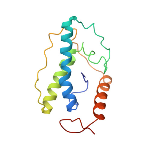
Crystallographic study of bacteriophage T4 endonuclease V, which is involved in the initial step of the pyrimidine dimer-specific excision repair pathway, has been carried out with respect to the wild-type and three different mutant enzymes. This enzyme catalyzes the cleavage of the N-glycosyl bond at the 5'-side of the pyrimidine dimer, and subsequently incises the phosphodiester bond at the apyrimidinic site through a beta-elimination reaction. The structure of the wild-type enzyme refined at 1.45 A resolution reveals the detailed molecular architecture. The enzyme is composed of a single compact domain classified as an all-alpha structure. The molecule is stabilized mainly by three hydrophobic cores, two of which include many aromatic side-chain interactions. The structure has a unique folding motif, where the amino-terminal segment penetrates between two major alpha-helices and prevents their direct contact, and it is incompatible with the close-packing category of helices for protein folding. The concave surface, covered with many positive charges, implies an interface for DNA binding. The glycosylase catalytic center, which comprises Glu23 and the surrounding basic residues Arg3, Arg22 and Arg26, lie in this basic surface. The crystal structures of the three active-site mutants, in which Glu23 was replaced by Gln(E23Q) and Asp (E23D), respectively, and Arg3 by Gln (R3Q), have been determined at atomic resolution. The backbone structures of the E23Q and R3Q mutants were almost identical with that of the wild-type, while the E23D mutation induces a small, but significant, change in the backbone structure, such as an increase of the central kink of the H1 helix at Pro25. In the catalytic center of the glycosylase, however, these three mutations do not generate notable movements of protein atoms, except for significant shifts of some bound water molecules. Thus, the structural differences between the wild-type and each mutant are confined to the remarkably small region around their replaced chemical groups. Combined with the biochemical studies and the difference circular dichroism measurements, these results allow us to conclude that the negatively charged carboxyl group of Glu23 is essential for the cleavage of the N-glycosyl bond, and that the positively charged guanidino group of Arg3 is crucial to bind the substrate, a DNA duplex containing a pyrimidine dimer. The amino terminal alpha-amino group is located at a position approximately 4.4 A away from the carboxyl group of Glu23. These structural features are generally consistent with the reaction scheme proposed by Dodson and co-workers.
Organizational Affiliation:
Protein Engineering Research Institute, Osaka, Japan.