


Crystal structure of Atlantic cod trypsin complexed with benzamidine
Iyaguchi, D., Toyota, E.To be published.
Experimental Data Snapshot
wwPDB Validation 3D Report Full Report

Entity ID: 1 | |||||
|---|---|---|---|---|---|
| Molecule | Chains | Sequence Length | Organism | Details | Image |
| Trypsin-1 | 220 | Gadus morhua | Mutation(s): 0 EC: 3.4.21.4 | 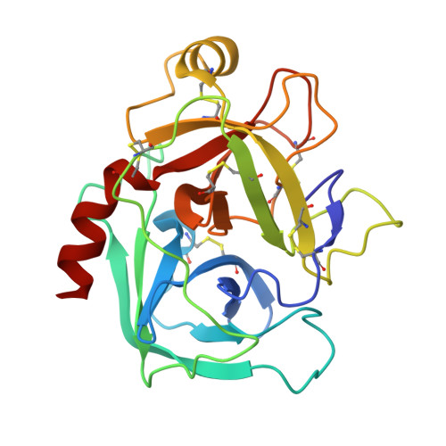 | |
UniProt | |||||
Find proteins for P16049 (Gadus morhua) Explore P16049 Go to UniProtKB: P16049 | |||||
Entity Groups | |||||
| Sequence Clusters | 30% Identity50% Identity70% Identity90% Identity95% Identity100% Identity | ||||
| UniProt Group | P16049 | ||||
Sequence AnnotationsExpand | |||||
| |||||
| Ligands 3 Unique | |||||
|---|---|---|---|---|---|
| ID | Chains | Name / Formula / InChI Key | 2D Diagram | 3D Interactions | |
| BEN Query on BEN | D [auth A] | BENZAMIDINE C7 H8 N2 PXXJHWLDUBFPOL-UHFFFAOYSA-N |  | ||
| CA Query on CA | B [auth A] | CALCIUM ION Ca BHPQYMZQTOCNFJ-UHFFFAOYSA-N |  | ||
| NA Query on NA | C [auth A] | SODIUM ION Na FKNQFGJONOIPTF-UHFFFAOYSA-N |  | ||
| Length ( Å ) | Angle ( ˚ ) |
|---|---|
| a = 47.213 | α = 90 |
| b = 59.184 | β = 90 |
| c = 63.736 | γ = 90 |
| Software Name | Purpose |
|---|---|
| CrystalClear | data collection |
| AMoRE | phasing |
| CNS | refinement |
| HKL-2000 | data reduction |
| HKL-2000 | data scaling |
RCSB PDB (citation) is hosted by
RCSB PDB is a member of the

