2E77

Crystal structure of L-lactate oxidase with pyruvate complex


Experimental Data Snapshot

  • Method: X-RAY DIFFRACTION
  • Resolution: 1.90 Å
  • R-Value Free: 0.227 
  • R-Value Work: 0.176 
  • R-Value Observed: 0.179 

wwPDB Validation   3D Report Full Report


Ligand Structure Quality Assessment 


This is version 2.0 of the entry. See complete history


Literature

Crystallographic study on the interaction of L-lactate oxidase with pyruvate at 1.9 Angstrom resolution.

Li, S.J.Umena, Y.Yorita, K.Matsuoka, T.Kita, A.Fukui, K.Morimoto, Y.

(2007) Biochem Biophys Res Commun 358: 1002-1007

  • DOI: https://doi.org/10.1016/j.bbrc.2007.05.021

  • PubMed Abstract: 

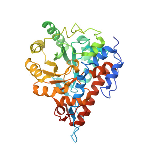
    L-Lactate oxidase (LOX) from Aerococcus viridans catalyzes the oxidation of L-lactate to pyruvate by the molecular oxygen and belongs to a large family of 2-hydroxy acid-dependent flavoenzymes. To investigate the interaction of LOX with pyruvate in structural details and understand the chemical mechanism of flavin-dependent L-lactate dehydrogenation, the LOX-pyruvate complex was crystallized and the crystal structure of the complex has been solved at a resolution of 1.90 Angstrom. One pyruvate molecule bound to the active site and located near N5 position of FMN for subunits, A, B, and D in the asymmetric unit, were identified. The pyruvate molecule is stabilized by the interaction of its carboxylate group with the side-chain atoms of Tyr40, Arg181, His265, and Arg268, and of its keto-oxygen atom with the side-chain atoms of Tyr146, Tyr215, and His265. The alpha-carbon of pyruvate is found to be 3.13 Angstrom from the N5 atom of FMN at an angle of 105.4 degrees from the flavin N5-N10 axis.


  • Organizational Affiliation

    Institute for Protein Research, Osaka University, Suita, Osaka 565-0871, Japan.


Macromolecules
Find similar proteins by:  (by identity cutoff)  |  3D Structure
Entity ID: 1
MoleculeChains Sequence LengthOrganismDetailsImage
Lactate oxidase
A, B, C, D
374Aerococcus viridansMutation(s): 0 
EC: 1.13.12.4
UniProt
Find proteins for Q44467 (Aerococcus viridans (strain ATCC 11563 / DSM 20340 / CCUG 4311 / JCM 20461 / NBRC 12219 / NCTC 8251 / M1))
Explore Q44467 
Go to UniProtKB:  Q44467
Entity Groups  
Sequence Clusters30% Identity50% Identity70% Identity90% Identity95% Identity100% Identity
UniProt GroupQ44467
Sequence Annotations
Expand
  • Reference Sequence
Experimental Data & Validation

Experimental Data

  • Method: X-RAY DIFFRACTION
  • Resolution: 1.90 Å
  • R-Value Free: 0.227 
  • R-Value Work: 0.176 
  • R-Value Observed: 0.179 
  • Space Group: C 1 2 1
Unit Cell:
Length ( Å )Angle ( ˚ )
a = 134.841α = 90
b = 118.566β = 121.07
c = 107.237γ = 90
Software Package:
Software NamePurpose
REFMACrefinement
ADSCdata collection
HKL-2000data reduction
SCALEPACKdata scaling
CNSphasing

Structure Validation

View Full Validation Report


Ligand Structure Quality Assessment 


Entry History 

Deposition Data

  • Released Date: 2007-11-27 
  • Deposition Author(s): Morimoto, Y.

Revision History  (Full details and data files)

  • Version 1.0: 2007-11-27
    Type: Initial release
  • Version 1.1: 2011-07-13
    Changes: Derived calculations, Version format compliance
  • Version 1.2: 2023-10-25
    Changes: Data collection, Database references, Derived calculations, Refinement description
  • Version 2.0: 2023-11-15
    Changes: Atomic model, Data collection