


Atomic resolution analysis of beta-1,3-xylanase catalytic module from Vibrio sp. AX-4
Sakaguchi, K., Kawamura, T., Watanabe, N., Kiyohara, M., Yamaguchi, K., Ito, M., Tanaka, I.To be published.
Experimental Data Snapshot
wwPDB Validation 3D Report Full Report

Entity ID: 1 | |||||
|---|---|---|---|---|---|
| Molecule | Chains | Sequence Length | Organism | Details | Image |
| beta-1,3-xylanase | 333 | Vibrio sp. AX-4 | Mutation(s): 0 EC: 3.2.1 | 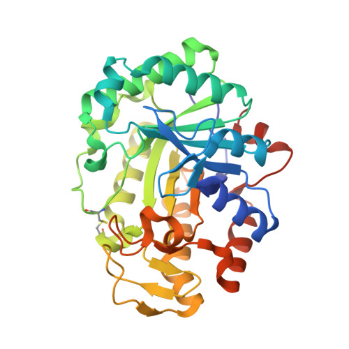 | |
UniProt | |||||
Find proteins for D5MP61 (Vibrio sp) Explore D5MP61 Go to UniProtKB: D5MP61 | |||||
Entity Groups | |||||
| Sequence Clusters | 30% Identity50% Identity70% Identity90% Identity95% Identity100% Identity | ||||
| UniProt Group | D5MP61 | ||||
Sequence AnnotationsExpand | |||||
| |||||
| Ligands 2 Unique | |||||
|---|---|---|---|---|---|
| ID | Chains | Name / Formula / InChI Key | 2D Diagram | 3D Interactions | |
| GOL Query on GOL | C [auth A], D [auth A], E [auth A] | GLYCEROL C3 H8 O3 PEDCQBHIVMGVHV-UHFFFAOYSA-N |  | ||
| MG Query on MG | B [auth A] | MAGNESIUM ION Mg JLVVSXFLKOJNIY-UHFFFAOYSA-N |  | ||
| Length ( Å ) | Angle ( ˚ ) |
|---|---|
| a = 51.873 | α = 90 |
| b = 75.654 | β = 90 |
| c = 82.119 | γ = 90 |
| Software Name | Purpose |
|---|---|
| REFMAC | refinement |
| HKL-2000 | data reduction |
| SCALEPACK | data scaling |
| SOLVE | phasing |
RCSB PDB (citation) is hosted by
RCSB PDB is a member of the

