2DCM

The Crystal Structure of S603A Mutated Prolyl Tripeptidyl Aminopeptidase Complexed with Substrate


Experimental Data Snapshot

  • Method: X-RAY DIFFRACTION
  • Resolution: 2.90 Å
  • R-Value Free: 0.263 
  • R-Value Work: 0.206 

wwPDB Validation   3D Report Full Report


Ligand Structure Quality Assessment 


This is version 1.4 of the entry. See complete history


Literature

Crystal Structure and Mechanism of Tripeptidyl Activity of Prolyl Tripeptidyl Aminopeptidase from Porphyromonas gingivalis

Ito, K.Nakajima, Y.Xu, Y.Yamada, N.Onohara, Y.Ito, T.Matsubara, F.Kabashima, T.Nakayama, K.Yoshimoto, T.

(2006) J Mol Biol 362: 228-240

  • DOI: https://doi.org/10.1016/j.jmb.2006.06.083
  • Primary Citation of Related Structures:  
    2D5L, 2DCM

  • PubMed Abstract: 

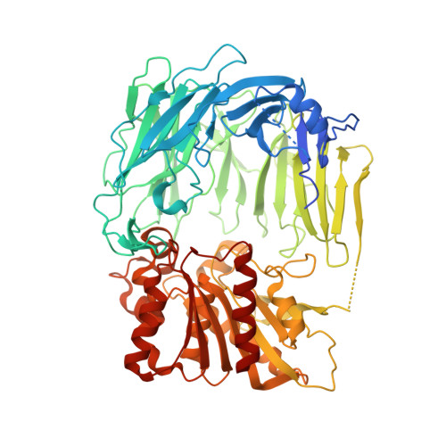
    The crystal structure of prolyl tripeptidyl aminopeptidase from Porphyromonas gingivalis was determined. Prolyl tripeptidyl aminopeptidase consists of beta-propeller and catalytic domains, and a large cavity between the domains; this structure is similar to dipeptidyl aminopeptidase IV. A catalytic triad (Ser603, His710, and Asp678) was located in the catalytic domain; this triad was virtually identical to that of the enzymes belonging to the prolyl oligopeptidase family. The structure of an inactive S603A mutant enzyme complexed with a substrate was also determined. The pyrrolidine ring of the proline residue appeared to fit into a hydrophobic pocket composed of Tyr604, Val629, Trp632, Tyr635, Tyr639, Val680, and Val681. There were characteristic differences in the residues of the beta-propeller domain, and these differences were related to the substrate specificity of tripeptidyl activity. The N-terminal amino group was recognized by salt bridges, with two carboxyl groups of Glu205 and Glu206 from a helix in dipeptidyl aminopeptidase IV. In prolyl tripeptidyl aminopeptidase, however, the Glu205 (located in the loop) and Glu636 were found to carry out this function. The loop structure provides sufficient space to accommodate three N-terminal residues (Xaa-Xaa-Pro) of substrates. This is the first report of the structure and substrate recognition mechanism of tripeptidyl peptidase.


  • Organizational Affiliation

    Gradute School of Biomedical Sciences, Nagasaki University 1-14 Bunkyo-machi, Nagasaki 852-8521, Japan.


Macromolecules
Find similar proteins by:  (by identity cutoff)  |  3D Structure
Entity ID: 1
MoleculeChains Sequence LengthOrganismDetailsImage
dipeptidyl aminopeptidase IV, putative706Porphyromonas gingivalis W83Mutation(s): 1 
Gene Names: PG1361
EC: 3.4.14
UniProt
Find proteins for Q7MUW6 (Porphyromonas gingivalis (strain ATCC BAA-308 / W83))
Explore Q7MUW6 
Go to UniProtKB:  Q7MUW6
Entity Groups  
Sequence Clusters30% Identity50% Identity70% Identity90% Identity95% Identity100% Identity
UniProt GroupQ7MUW6
Sequence Annotations
Expand
  • Reference Sequence
Small Molecules
Ligands 1 Unique
IDChains Name / Formula / InChI Key2D Diagram3D Interactions
GA0
Query on GA0

Download Ideal Coordinates CCD File 
B [auth A]GLYCYLALANYL-N-2-NAPHTHYL-L-PROLINEAMIDE
C20 H24 N4 O3
KCELZXZDIUJGNM-GUYCJALGSA-N
Experimental Data & Validation

Experimental Data

  • Method: X-RAY DIFFRACTION
  • Resolution: 2.90 Å
  • R-Value Free: 0.263 
  • R-Value Work: 0.206 
  • Space Group: P 63 2 2
Unit Cell:
Length ( Å )Angle ( ˚ )
a = 149.453α = 90
b = 149.453β = 90
c = 159.644γ = 120
Software Package:
Software NamePurpose
HKL-2000data collection
SCALEPACKdata scaling
CNSrefinement
HKL-2000data reduction
CNSphasing

Structure Validation

View Full Validation Report


Ligand Structure Quality Assessment 


Entry History 

Deposition Data

Revision History  (Full details and data files)

  • Version 1.0: 2006-09-19
    Type: Initial release
  • Version 1.1: 2008-04-30
    Changes: Version format compliance
  • Version 1.2: 2011-07-13
    Changes: Derived calculations, Source and taxonomy, Version format compliance
  • Version 1.3: 2021-11-10
    Changes: Database references, Derived calculations
  • Version 1.4: 2023-10-25
    Changes: Data collection, Refinement description