Role of active site water molecules and substrate hydroxyl groups in oxygen activation by cytochrome P450 158A2: a new mechanism of proton transfer
Zhao, B., Guengerich, F.P., Voehler, M., Waterman, M.R.(2005) J Biol Chem 280: 42188-42197
- PubMed: 16239228
- DOI: https://doi.org/10.1074/jbc.M509220200
- Primary Citation of Related Structures:
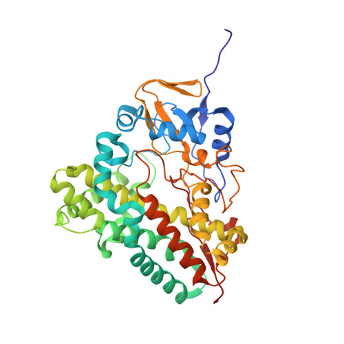
2D09, 2D0E - PubMed Abstract:
From the x-ray crystal structure of CYP158A2 (Zhao, B., Guengerich, F. P., Bellamine, A., Lamb, D. C., Izumikawa, M., Lei, L., Podust, L. M., Sundaramoorthy, M., Reddy, L. M., Kelly, S. L., Kalaitzis, J. A., Stec, D., Voehler, M., Falck, J. R., Moore, B. S., Shimada, T., and Waterman, M. R. (2005) J. Biol. Chem. 280, 11599-11607), one of 18 cytochrome P450 (CYP) genes in the actinomycete Streptomyces coelicolor, ordered active site water molecules (WAT505, WAT600, and WAT640), and hydroxyl groups of the substrate flaviolin were proposed to participate in proton transfer and oxygen cleavage in this monooxygenase. To probe their roles in catalysis, we have studied the crystal structures of a substrate analogue (2-hydroxy-1,4-naphthoquinone) complex with ferric CYP158A2 (2.15 A) and the flaviolin ferrous dioxygen-bound CYP158A2 complex (1.8 A). Catalytic activity toward 2-hydroxy-1,4-naphthoquinone was approximately 70-fold less than with flaviolin. In the ferrous dioxygen-bound flaviolin complex, the three water molecules in the ferric flaviolin complex still occupy the same positions and form hydrogen bonds to the distal dioxygen atom. These findings suggest that CYP158A2 utilizes substrate hydroxyl groups to stabilize active site water and further assist in the iron-linked dioxygen activation. A continuous hydrogen-bonded water network connecting the active site to the protein surface (bulk solvent) not present in the other two ferrous dioxygen-bound P450 structures (CYP101A1/P450cam and CYP107A1/P450eryF) is proposed to participate in the proton-delivery cascade, leading to dioxygen bond scission. This ferrous-dioxygen structure suggests two classes of P450s based on the pathway of proton transfer, one using the highly conserved threonine in the I-helix (CYP101A1) and the other requiring hydroxyl groups of the substrate molecules either directly transferring protons (CYP107A1) or stabilizing a water pathway for proton transfer (CYP158A2).
Organizational Affiliation:
Department of Biochemistry, Centers for Structural Biology, Vanderbilt University School of Medicine, Nashville, Tennessee 37235, USA. bin.zhao@vanderbilt.edu