Structure of RadB recombinase from a hyperthermophilic archaeon, Thermococcus kodakaraensis KOD1: an implication for the formation of a near-7-fold helical assembly
Akiba, T., Ishii, N., Rashid, N., Morikawa, M., Imanaka, T., Harata, K.(2005) Nucleic Acids Res 33: 3412-3423
- PubMed: 15956102
- DOI: https://doi.org/10.1093/nar/gki662
- Primary Citation of Related Structures:
2CVF, 2CVH - PubMed Abstract:
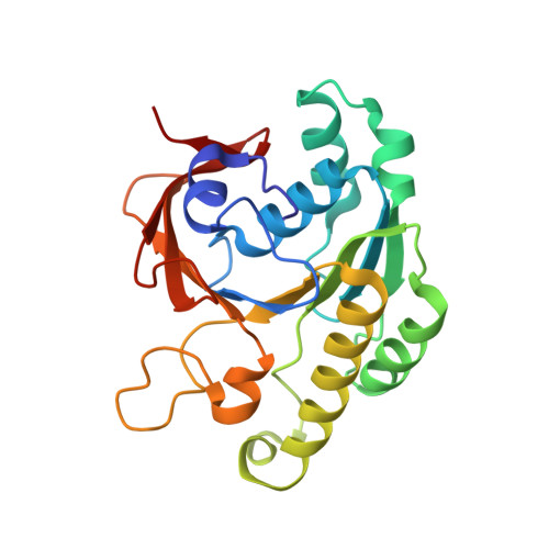
The X-ray crystal structure of RadB from Thermococcus kodakaraensis KOD1, an archaeal homologue of the RecA/Rad51 family proteins, have been determined in two crystal forms. The structure represents the core ATPase domain of the RecA/Rad51 proteins. Two independent molecules in the type 1 crystal were roughly related by 7-fold screw symmetry whereas non-crystallographic 2-fold symmetry was observed in the type 2 crystal. The dimer structure in the type 1 crystal is extended to construct a helical assembly, which resembles the filamentous structures reported for other RecA/Rad51 proteins. The molecular interface in the type 1 dimer is formed by facing a basic surface patch of one monomer to an acidic one of the other. The empty ATP binding pocket is located at the interface and barely concealed from the outside similarly to that in the active form of the RecA filament. The model assembly has a positively charged belt on one surface bordering the helical groove suitable for facile binding of DNA. Electron microscopy has revealed that, in the absence of ATP and DNA, RadB forms a filament with a similar diameter to that of the hypothetical assembly, although its helical properties were not confirmed.
Organizational Affiliation:
Biological Information Research Center, National Institute of Advanced Industrial Science and Technology (AIST) Tsukuba Central 6, 1-1-1 Higashi, Tsukuba, Ibaraki 305-8566, Japan.